. 2007 Aug;19(8):2544-56.
doi: 10.1105/tpc.107.051797. Epub 2007 Aug 10.

Genetic and molecular interactions between BELL1 and MADS box factors support ovule development in Arabidopsis

Affiliations

Genetic and molecular interactions between BELL1 and MADS box factors support ovule development in Arabidopsis

Vittoria Brambilla et al. Plant Cell. 2007 Aug.

Abstract

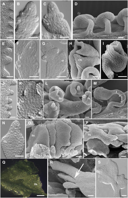
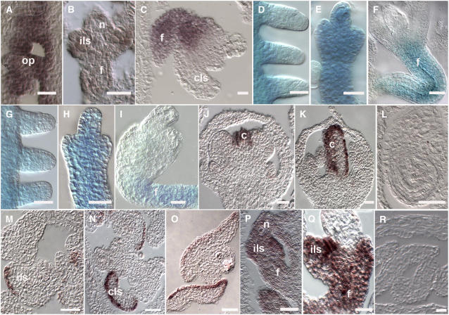
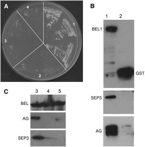
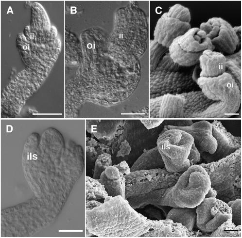
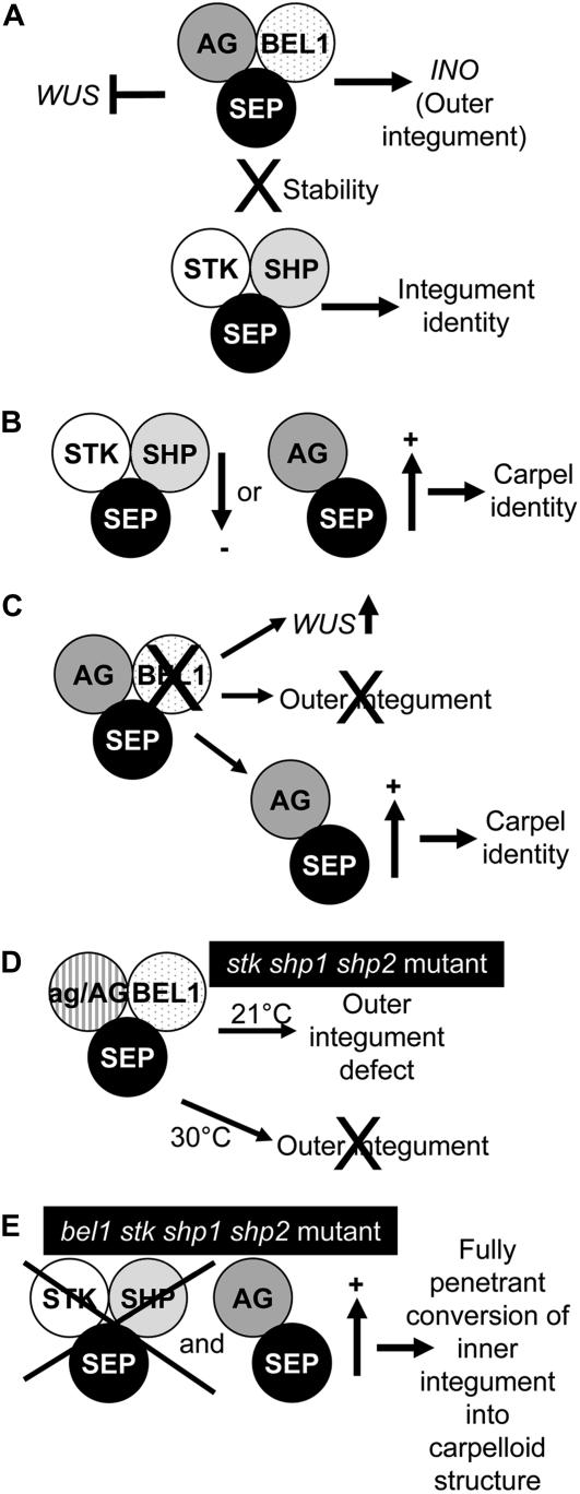
In Arabidopsis thaliana and many other plant species, ovules arise from carpel tissue as new meristematic formations. Cell fate in proliferating ovule primordia is specified by particular ovule identity factors, such as the homeodomain factor BELL1 (BEL1) and MADS box family members SEEDSTICK (STK), SHATTERPROOF1 (SHP1), SHP2, and AGAMOUS. Both in the bel1 mutant and the stk shp1 shp2 triple mutant, integuments are transformed into carpelloid structures. Combining these mutants in a bel1 stk shp1 shp2 quadruple mutant, we showed that the bel1 phenotype is significantly enhanced. We also demonstrate that ovule differentiation requires the regulation of the stem cell maintenance gene WUSCHEL, repression of which is predominantly maintained by BEL1 during ovule development. Based on yeast three-hybrid assays and genetic data, we show that BEL1 interacts with the ovule identity MADS box factors when they dimerize with SEPALLATA proteins. We propose a model for ovule development that explains how the balance between carpel identity activity and ovule identity activity is established by a MADS box homeodomain protein complex.


Figures

Figure 1.
Figure 1.
Ovule Development in Wild-Type and Mutant Plants. (A) to (D) Ovule development in wild-type plants. (A) to (C) Differential interference contrast (DIC) microscopy images. (A) Ovule primordia arise from the placenta at stage 9 of flower development. (B) The nucellus and funiculus differentiate at stage 11. Inner and outer integument primordia are also visible. (C) The outer integument grows asymmetrically. (D) Megagametogenesis is initiated. Scanning electron microscopy image of wild-type mature ovules. (E) to (I) Ovule development in the stk shp1 shp2 triple mutant. (E) to (G) DIC microscopy images. (E) Ovule primordia arise from the placenta at stage 9 of flower development. (F) Inner and outer integuments develop normally until stage 11. (G) Integument development is affected starting from stage 12. (H) and (I) Scanning electron microscopy images of mature ovules. (H) Ovules are converted in carpel-like structures. The funiculus is still visible, while integuments develop in a carpel-like structure. (I) In some ovules, integuments don't show homeotic conversion; nevertheless, the growth is affected. The outer integument is only partially developed, and it doesn't overgrow the inner integument and the nucellus. (J) to (L) Ovule development in the bel1 mutant. (J) and (K) DIC microscopy images. (J) Ovule primordia arising from the placenta. (K) At stage 11, a single and thick integument-like structure differentiates in the chalaza region. (L) Scanning electron microscopy image of bel1-1 ovules. The integument-like structures expand at the base of the nucellus. (M) Scanning electron microscopy image of the bel1 stk mutant ovule. The ovule phenotype is additive with respect to the single mutants. A thicker and longer funiculus than the wild type is visible. Integument-like structures develop in the chalaza region. (N) to (S) Ovule development of the bel1 stk shp1 shp2 mutant. (N) and (S) DIC microscopy images. (O), (P), and (R) Scanning electron microscopy images. (N) At stage 11, integument development resembles the bel1 single mutant. A thick integument-like structure is visible. (O) and (P) Integuments expand, forming leaf-like structures. This phenotype is fully penetrant. (Q) Stereomicroscopy image of an ovule converted in a green leaf-like structure. (R) The arrow indicates lateral outgrowth from the chalaza region. (S) In some cases, secondary mutant ovule-like structures develop from the funiculus. n, nucellus; ii, inner integument; oi, outer integument; cls, carpel-like structures; ils, integument-like structures; lls, leaf-like structures; f, funiculus. Bars = 25 μM.
Figure 2.
Figure 2.
Gene Expression in Wild-Type and in bel1, stk shp1 shp2, and bel1 stk shp1 shp2 Mutant Ovules. (A) to (C) STK expression profile in bel1-1 mutant ovules as detected by in situ hybridization. (A) STK mRNA is detected in the placenta and in ovule primordia. (B) At stage 12, STK is still expressed in the funiculus and in the integument-like structure that develops from the chalaza region. (C) STK expression is not detected in the carpel-like structures developing from the chalaza but is restricted to the funiculus. (D) to (F) Expression of the STK:GUS reporter gene in the bel1-1 mutant. (D) GUS activity is detected in the placenta and in ovule primordia. (E) Until stage 12, GUS activity is detected throughout the ovule. (F) During late stage 12, GUS activity is restricted to the funiculus. (G) to (I) Expression of the STK:GUS reporter gene in the bel stk shp1 shp2 mutant ovules. (G) and (H) GUS activity is detected in placenta and in developing ovules until stage 12. (I) At later stages, GUS activity is detectable only in the funiculus. (J) to (O) CRC expression profile as detected by in situ hybridization. (J) to (L) Wild-type flower. (J) At stage 7 of flower development, CRC is transcribed in developing carpels. (K) During carpel development, CRC expression is restricted to the epidermal cell layers. (L) CRC expression is not detected in mature ovules. (M) bel1 ovules. CRC mRNA is detectable in the integument-like structures that develop in the chalaza region. The expression pattern resembles the situation in wild-type carpels, where only particular cell layers show the hybridization signal. (N) stk shp1 shp2 ovules. CRC is expressed in the carpel-like structures that develop in mature mutant ovules. (O) be1l stk shp1 shp2 ovules. CRC mRNA is detectable in the converted structure developing in the chalaza region. (P) to (R) WUS expression profile in bel1 stk shp1 shp2 ovules as detected by in situ hybridization. (P) Stage 11 of flower development. In the quadruple mutant, WUS mRNA is present in all the ovules. (Q) At later stages, WUS is strongly expressed in the integument-like structure and in the funiculus. (R) WUS mRNA is not detectable in the carpel-like structures at later stages of development. f, funiculus; c, carpel; cls, carpel-like structure; ils, integument-like structures; op, ovule primordia; n, nucellus. Bars = 25 μM.
Figure 3.
Figure 3.
Interaction Assays to Test for BEL1, SEP, and AG Protein Interactions. (A) Yeast three-hybrid assays using selective media without His. (1) Three independent yeast transformants cotransformed with pBD-AG, pTFT-SEP1, and pAD-BEL1; (2) to (4) independent yeast transformants using different empty vectors as control. (2) pAD-empty; (3) pTFT-empty; (4) pBD-empty. (B) and (C) In vitro pull-down assays to test BEL1, SEP3, and AG protein interactions. Extracts of Escherichia coli cells expressing BEL1-GST, AG-MBP, SEP3-TRX, and only the GST tag were used to confirm the physical interactions occurring among these three transcription factors. Bound proteins were analyzed by SDS-PAGE and visualized by immunoblotting using antibodies against GST (top panels), MBP (middle panels), and TRX (bottom panels). First, we allowed the formation of the AG-MAL/SEP3-TRX heterodimer; subsequently, this complex was incubated with BEL-GST protein linked to glutathione-agarose beads (lane 1 in [B] and lanes 1 to 3 in [C]). As a negative control, the AG-MAL/SEP3-TRX heterodimer was also applied to GST bound to glutathione-agarose beads. AG and SEP3 are coimmunoprecipitated with BEL1 (lane 1 in [B] and [C]), but they do not interact with the GST tag (lane 2 in [B]). Weak interactions occur between BEL1 and SEP3 (cf. lanes 1 and 2 of [C]) or BEL1 and AG (cf. lanes 1 and 3 of [C]). Proteins are indicated close to the corresponding bands, and in each lane, equal amounts of protein were loaded.
Figure 4.
Figure 4.
Ovule Morphology in the ag/AG stk shp1 shp2 Mutant. (A) to (C) ag/AG stk shp1 shp2 mutant grown at 21°C. (A) and (B) DIC microscopy images. (A) Integument development proceeds like in the wild-type plant until stage 10. Inner and outer integument primordia emerge from the chalaza. (B) At later stages, integument development is arrested in all the ovules. (C) Scanning electron microscopy image. Outer and inner integuments do not overgrow the nucellus. (D) and (E) ag/AG stk shp1 shp2 mutant grown at 30°C. (D) DIC microscopy image. An integument-like structure develops at the base of the nucellus resembling bel1 ovules. (E) Scanning electron microscopy image of ag/AG stk shp1 shp1 ovules that resemble bel1 ovules. ii, inner integument; oi, outer integument; ils, integument-like structure. Bars = 25 μM.
Figure 5.
Figure 5.
A Model for Integument Identity Determination and Development. (A) In wild-type ovules, the BEL1 protein interacts with the AG-SEP dimer to repress WUS in the chalaza and to regulate outer integument development. This complex is stabilized by the ovule identity complex. (B) When the ovule identity complex is reduced or the AG-SEP dimer is increased, integuments acquire carpel identity. (C) In the bel1 mutant, the amount of AG-SEP dimer is increased. In this case, WUS is ectopically expressed in the chalaza and the outer integument does not develop. Moreover, the inner integument develops like a carpel structure. (D) In the stk shp1 shp2 ag/AG mutant, there is still enough AG-BEL-SEP complex to regulate inner and outer integument development. However, at higher temperatures, this complex is very unstable due to the absence of the ovule identity complex, and the outer integument does not develop. (E) In the stk shp1 shp2 bel quadruple mutant, only the SEP-AG dimer is available, resulting in the conversion of the inner integument into a carpelloid structure.

Similar articles

Cited by

References

    1. Alvarez, J., and Smyth, D.R. (1999). CRABS CLAW and SPATULA, two Arabidopsis genes that control carpel development in parallel with AGAMOUS. Development 126 2377–2386. - PubMed
    1. Baker, S.C., Robinson-Beers, K., Villanueva, J.M., Gaiser, J.C., and Gasser, C.S. (1997). Interactions among genes regulating ovule development in Arabidopsis thaliana. Genetics 145 1109–1124. - PMC - PubMed
    1. Balasubramanian, S., and Schneitz, K. (2002). NOZZLE links proximal-distal and adaxial-abaxial pattern formation during ovule development in Arabidopsis thaliana. Development 129 4291–4300. - PubMed
    1. Battaglia, R., Brambilla, V., Colombo, L., Stuitje, A.R., and Kater, M.M. (2006). Functional analysis of MADS-box genes controlling ovule development in Arabidopsis using the ethanol-inducible alc gene-expression system. Mech. Dev. 123 267–276. - PubMed
    1. Bellaoui, M., Pidkowich, M.S., Samach, A., Kushalappa, K., Kohalmi, S.E., Modrusan, Z., Crosby, W.L., and Haughn, G.W. (2001). The Arabidopsis BELL1 and KNOX TALE homeodomain proteins interact through a domain conserved between plants and animals. Plant Cell 13 2455–2470. - PMC - PubMed

Publication types

MeSH terms