. 2007 Oct;145(2):437-49.
doi: 10.1104/pp.107.097089. Epub 2007 Aug 10.

Medicago truncatula as a model for nonhost resistance in legume-parasitic plant interactions

Affiliations

Medicago truncatula as a model for nonhost resistance in legume-parasitic plant interactions

M Dolores Lozano-Baena et al. Plant Physiol. 2007 Oct.

Abstract

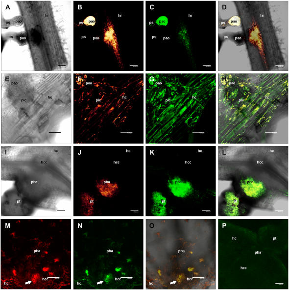
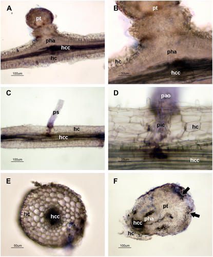
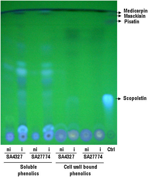
Crenate broomrape (Orobanche crenata) is a root parasitic weed that represents a major constraint for grain legume production in Mediterranean and West Asian countries. Medicago truncatula has emerged as an important model plant species for structural and functional genomics. The close phylogenic relationship of M. truncatula with crop legumes increases its value as a resource for understanding resistance against Orobanche spp. Different cytological methods were used to study the mechanisms of resistance against crenate broomrape of two accessions of M. truncatula, showing early and late acting resistance. In the early resistance accession (SA27774) we found that the parasite died before a tubercle had formed. In the late resistance accession (SA4327) the parasite became attached without apparent problems to the host roots but most of the established tubercles turned dark and died before emergence. The results suggest that there are defensive mechanisms acting in both accessions but with a time gap that is crucial for a higher success avoiding parasite infection.


Figures

Figure 1.
Figure 1.
Sections stained with TBO. A, Cross section of a successful crenate broomrape seedling penetration on SA4087 accession of M. truncatula. B, Detail of A showing the central cylinder and host xylem vessels in contact with parasite cells. Some parasite vessels begin to develop connecting with the host xylem vessels. C, Cross section of a successful crenate broomrape seedling penetration on SA4327 accession of M. truncatula. D, Detail of C showing the central cylinder and host xylem vessels in contact with parasite cells. E, Cross section of an unsuccessful crenate broomrape seedling penetration on SA27774 accession of M. truncatula. F, Detail of E showing the thickening of host xylem walls (arrows) in contact with parasite cells and accumulation of a dark stained substance (arrowhead). ps, Parasite seedling; pic, parasite intrusive cells; hc, host cortex; hcc, host central cylinder; hx, host xylem vessels; px, parasite xylem vessels. [See online article for color version of this figure.]
Figure 2.
Figure 2.
Sections stained with AGS. A, Cross section of an unsuccessful crenate broomrape seedling penetration on SA27774 accession of M. truncatula. B, The same section observed under UV excitation (340–380 nm) showing an intense fluorescence in host cells in contact with parasite tissues (arrows). C, Overlay of A and B showing the localization of the fluorescent cells. D, Detail of A showing accumulation of substances (noncarbohydrates) inside host xylem vessels (arrows) and thickening of xylem vessels in contact with parasite cells (arrowhead). E, The same section as D observed under UV excitation (340–380 nm) and showing blue fluorescence from the thickened xylem cell walls (arrowhead). F, The same section as D observed under polarized light showing no changes in the cell walls birefringence of thickened xylem vessels. G, Cross section of a successful established crenate broomrape tubercle on SA4087 accession. H, Cross section of a darkened established crenate broomrape tubercle on SA4327 accession. I, The same section as H observed under polarized light. J, Detail of a cross section of a darkened established crenate broomrape tubercle on SA4327 accession showing accumulation of a dark brown substance inside parasite xylem vessels and the apoplast of the haustorium (arrow). K, The same section as J observed under UV excitation (340–380 nm) and showing quenched autofluorescence from the vessels covered by the dark brown substance (arrow). ps, Parasite seedling; pic, parasite intrusive cells; hc, host cortex; hcc, host central cylinder; hx, host xylem vessels; pha, parasite haustorium; pt, parasite tubercle; px, parasite xylem vessels.
Figure 3.
Figure 3.
Sections stained with phloroglucinol-HCl. A, Cross section of an unsuccessful crenate broomrape seedling penetration on SA27774 accession of M. truncatula. B, The same section observed under UV excitation (340–380 nm). C, Detail of A showing accumulation of substances (polyphenols) inside host xylem vessels and thickening of their cell walls (arrows) compared with normal xylem vessels stained with the dye (arrowhead). D, The same section as C observed under UV excitation (340–380 nm) and showing blue fluorescence corresponding to suberin from endodermal cells, and the quenched fluorescence from normal lignified xylem walls (arrowhead) and thickened xylem walls (arrows). E, Cross section of a successful crenate broomrape seedling penetration on SA4087 accession of M. truncatula. F, Detail of E showing the normal staining of host xylem walls. ps, Parasite seedling; pic, parasite intrusive cells; hc, host cortex; hcc, host central cylinder; hx, host xylem vessels. [See online article for color version of this figure.]
Figure 4.
Figure 4.
Sections stained with aniline blue fluorochrome. A, General view of a cross section of an incompatible interaction on SA27774 accession of M. truncatula observed under bright field. B, The same section as A observed under UV excitation (340–380 nm). C, Detail of B showing callose depositions (arrow) in cell walls from the host cortex in contact with parasite cells. D, The same section as C showing callose depositions (arrow) in cell walls from the host central cylinder. E, Cross section of a compatible interaction on SA4087 accession of M. truncatula observed under bright field. F, The same section as E observed under UV excitation (340–380 nm). No presence of callose is detected. pt, Parasite tubercle; pic, parasite intrusive cells; hc, host cortex; hcc, host central cylinder.
Figure 5.
Figure 5.
Hand cut fresh sections for fluorescence observation. A, Longitudinal section of an uninfected M. truncatula root observed under bright field. B, The same section as A observed under UV excitation (340–380 nm). C, Longitudinal section of a successful established crenate broomrape tubercle on SA4327 accession of M. truncatula observed under bright field. D, The same section as C observed under UV excitation (340–380 nm). E, Longitudinal section of an unsuccessful crenate broomrape seedling penetration on SA27774 accession of M. truncatula observed under bright field. F, The same section as E observed under UV excitation (340–380 nm) and showing a strong fluorescence from host cells in contact with parasite tissues (arrows). G, Cross section of a darkened crenate broomrape tubercle on SA4327 accession observed under bright field. H, The same section as G observed under UV excitation (340–380 nm) and showing a strong fluorescence from host cells in contact with parasite tissues (arrows). hc, Host cortex; hcc, host central cylinder; pha, parasite haustorium; pt, parasite tubercle; pao, parasite attachment organ; pic, parasite intrusive cells.
Figure 6.
Figure 6.
Localization of phenolic compounds using confocal laser microscopy. The images are single optical sections in B, C, F, G, J, K, M, N, and P. Figures B, F, J, and M correspond to emission spectra collected within the red channel (590–670 nm). Figures C, G, K, N, and P correspond to emission spectra collected within the green channel (515–590 nm). A, Incompatible interaction of crenate broomrape on SA27774 accession of M. truncatula (transmission). A darkening of the attachment organ and root tissues can be observed. B, Optical section of A showing intense fluorescence (red channel) in the host central cylinder and in the youngest attachment organ. C, The same section as B through the green channel. D, Overlay of A, B, and C showing the localization of the fluorescence in tissues. E, Detail of an unsuccessful crenate broomrape seedling penetration on SA27774 accession (transmission). F, Optical section of E showing intense fluorescence (red channel) in the host cells and in the parasite intrusive cells. G, The same section as F through the green channel. H, Overlay of E, F, and G showing the localization of the fluorescence in tissues. I, Darkened crenate broomrape tubercle on SA4327 accession of M. truncatula (transmission). J, Optical section of I showing intense fluorescence (red channel) in the haustorium and distal parts of the tubercle. K, The same section as J through the green channel. L, Overlay of I, J, and K showing the localization of the fluorescence in tissues. M, Optical section of a darkened crenate broomrape tubercle on SA4327 accession showing intense fluorescence (red channel) in the host xylem vessels (arrow). N, The same section as M through the green channel. O, Overlay of M and N with a transmission image showing the localization of the fluorescence in tissues. P, Optical section of a normal crenate broomrape tubercle on SA4327 accession. No fluorescence (green channel) can be detected. ps, Parasite seedling; pao, parasite attachment organ; hr, host root; pic, parasite intrusive cells; hc, host cortex; hcc, host central cylinder; pha, parasite haustorium; pt, parasite tubercle. Scale bar: 80 μm in A to D, I to L, and P; 40 μm in E to H and M to O.
Figure 7.
Figure 7.
Hand cut fresh sections stained for cell viability. A, Longitudinal section of a normal crenate broomrape tubercle on SA4327 accession of M. truncatula. B, Detail of A showing the absence of stained cells in host and parasite tissues. C, Longitudinal section of an unsuccessful crenate broomrape seedling penetration on SA27774 accession of M. truncatula. D, Detail of C showing stained cells corresponding to the parasite intrusive cells and the attachment organ, indicating their loss of viability. E, Cross section of an uninfected M. truncatula root showing the absence of stained cells. F, Cross section of a darkened crenate broomrape tubercle on SA4327 accession showing stained cells (arrows) in the distal part of the tubercle. pt, Parasite tubercle; pha, haustorium; hc, host cortex; hcc, host central cylinder; ps, parasite seedling; pao, parasite attachment organ; pic, parasite intrusive cells.
Figure 8.
Figure 8.
TLC plate of the methanolic extract from root samples of crenate broomrape inoculated (i) and noninoculated (ni) M. truncatula plants. Ctrl, Control with standard phytoalexins (scopoletin, medicarpin, pisatin, and maackiain).

Similar articles

Cited by

References

    1. Antonova TS, Ter Borg SJ (1996) The role of peroxidase in the resistance of sunflower against Orobanche cumana in Russia. Weed Res 36 113–121
    1. Baayen RP, Ouellette GB, Rioux D (1996) Compartmentalization of decay in carnations resistant to Fusarium oxysporum f. sp. dianthi. Phytopathology 86 1018–1031
    1. Blondon F, Marie D, Brown S, Kondorosi A (1994) Genome size and base composition in Medicago sativa and M. truncatula species. Genome 37 264–275 - PubMed
    1. Bordallo JJ, Lopez-Llorca LV, Jansson HB, Salinas J, Persmark L, Asensio L (2002) Colonization of plant roots by egg-parasitic and nematode-trapping fungi. New Phytol 154 491–499 - PubMed
    1. Cachinero JM, Hervás A, Jiménez-Díaz RM, Tena M (2002) Plant defence reactions against fusarium wilt in chickpea induced by incompatible race 0 of Fusarium oxysporum f.sp. ciceris and nonhost isolates of F. oxysporum. Plant Pathol 51 765–776

Publication types

LinkOut - more resources