. 2007 Sep 4;104(36):14182-8.
doi: 10.1073/pnas.0705994104. Epub 2007 Aug 10.

A pathogenicity island replicon in Staphylococcus aureus replicates as an unstable plasmid

Affiliations

A pathogenicity island replicon in Staphylococcus aureus replicates as an unstable plasmid

Carles Ubeda et al. Proc Natl Acad Sci U S A. .

Abstract

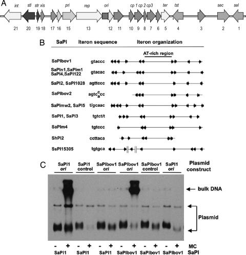
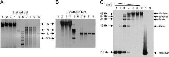
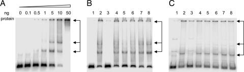
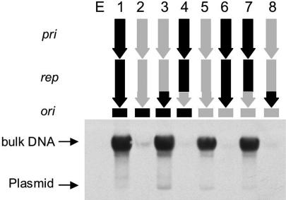
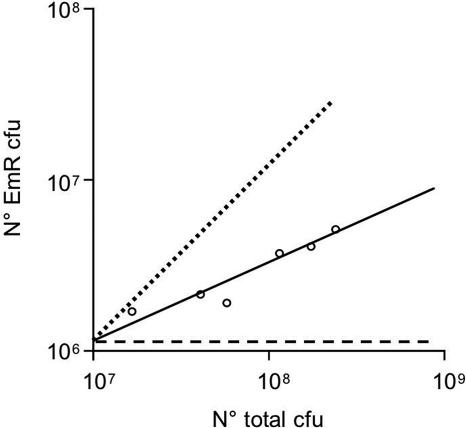
The SaPIs are 14- to 17-kb mobile pathogenicity islands in staphylococci that carry genes for superantigen toxins and other virulence factors and are responsible for the toxic shock syndrome and other superantigen-related diseases. They reside at specific chromosomal sites and are induced by certain bacteriophages to initiate an excision-replication-packaging program, resulting in their incorporation into small infective phage-like particles. These are responsible for very high transfer frequencies that often equal and sometimes exceed the plaque-forming titer of the inducing phage. The ability of the SaPIs to replicate autonomously defines them as individual replicons and, like other prokaryotic replicons, they possess replicon-specific initiation functions. In this paper, we report identification of the SaPI replication origin (ori) and replication initiation protein (Rep), which has helicase as well as initiation activity. The SaPI oris are binding sites for the respective Rep proteins and consist of multiple oligonucleotide repeats in two sets, flanking an AT-rich region that may be the site of initial melting. Plasmids containing the rep-ori complex plus an additional gene, pri, can replicate autonomously in Staphylococcus aureus but are very unstable, probably because of defective segregation.


Conflict of interest statement

The authors declare no conflict of interest.

Figures

Fig. 1.
Fig. 1.
Identification of the SaPI replication origins. (A) Map of SaPIbov1. Arrows represent the localization and orientation of the different ORFs, with labels indicating known gene functions. No functions have been identified for ORFs 3, 6, 10, 11, 12, 16, and 17, which have no significant matches in the database. (B) Comparative map of the replication origins of several SaPIs. The iterons are represented by arrows, and their sequences are shown at left. Note that there are always two sets of iterons flanking an AT-rich region, which could be the melting site. (C) The sequences containing the iterons of SaPIbov1 or SaPI1 were cloned into the thermosensitive plasmid pBT2. Sequences not containing iterons of each island were cloned as controls. The resulting plasmids were introduced into 80α lysogens carrying SaPI1 or SaPIbov1. Bacterial cultures of the different strains were exposed to MC, then incubated in broth at 43°C (restrictive temperature for pBT2 replication). Samples were removed 90 min after MC induction. Standard minilysates were prepared, separated by agarose gel electrophoresis, and blotted by using a pBT2-specific probe.
Fig. 2.
Fig. 2.
Replication products of SaPIbov1 pri-rep-ori subclone. (A) Total DNA from bacterial cultures was extracted and separated by agarose gel electrophoresis (lanes 1–4) or was initially digested with EcoRI, which cuts once in the plasmid (lanes 7–9). Lane 1, RN4220; lanes 2–4 and 7–9 correspond to three EmR transformants obtained in RN4220 with the SaPIbov1 pri-rep-ori subclone (pRN9211); lane 5 corresponds to the plasmid pRN9211 extracted from E. coli strain DH5α; lane 10, same plasmid DNA as lane 5 previously digested with EcoRI; lane 6, DNA ladder. (B) The same gel probed with a pRN9211-specific probe. The locations of multimers (M), nicked circular monomers (N), linear monomers (L), and supercoiled monomers (SC) are indicated. (C) Whole-cell DNA isolated from one pri-rep-ori transformant, digested with diminishing amounts of EcoRI (lanes 1–6) or undigested (lane 7), was separated by agarose gel electrophoresis, then Southern blotted as in B. Lane 8, plasmid pRN9211 extracted from E. coli and digested with EcoRI; lane 9, RN4220 DNA digested with EcoRI.
Fig. 3.
Fig. 3.
Specific binding of Rep protein to the cognate SaPI replication origin. (A) Gel mobility-shift assay by using SaPIbov1-ori as probe and increasing amounts of purified SaPIbov1 Rep protein. The different DNA–protein complexes are indicated by arrows. (B) Competition assays by using 5 ng of purified SaPIbov1-Rep protein, labeled SaPIbov1-ori DNA, and 10-fold excess of unlabeled DNA (lanes 3–8). SaPIbov1-ori DNA (lane 3), nonspecific DNA (lane 4), SaPI2-ori DNA (lane 5), SaPIn1-ori DNA (lane 6), SaPI1-ori DNA (lane 7), and SaPIm4-ori DNA (lane 8) were used as competitor unlabeled DNA. Lane 1 is a negative control without protein and unlabeled DNA. Lane 2 is a positive control without unlabeled DNA. (C) Competition assays by using 50 ng of purified SaPI1 Rep protein, labeled SaPI1-ori DNA, and 10-fold excess of unlabeled DNA (lanes 3–8). SaPI1-ori DNA (lane 3), nonspecific DNA (lane 4), SaPIm4-ori DNA (lane 5), SaPIbov1-ori DNA (lane 6), SaPIbov2-ori DNA (lane 7), and SaPIn1-ori DNA (lane 8) were used as competitor unlabeled DNA. Lane 1 is a negative control without protein and unlabeled DNA. Lane 2 is a positive control without unlabeled DNA.
Fig. 4.
Fig. 4.
Rep-ori replication specificity. Cultures of RN4220 strains containing the pri-rep-ori modules of SaPIbov1 and SaPI2 and chimeric constructs as shown were grown on CY broth at 43°C until OD540 = 0.6. Samples were removed and used to prepare minilysates. Lysates were separated by agarose gel electrophoresis and probed with a probe specific for the carrier plasmid, pRN9220. Above the blot is shown the composition of the pri-rep-ori module of each of the plasmids tested. Black indicates segments derived from SaPIbov1, gray, from SaPI2. E indicates empty vector.
Fig. 5.
Fig. 5.
SaPIbov1 pri-rep-ori subclone stability. Bacteria RN4220 containing the SaPIbov1 pri-rep-ori subclone grown on selective medium were grown on CY broth without antibiotic. Samples were taken every 30 min during 3 h and plated on GL plates with or without Em. The graph shows the number of EmR colonies obtained in relation to the total number of colonies obtained during the 3 h (solid line). The dashed line represents a plasmid that does not replicate. The dotted line represents a stable plasmid.
Fig. 6.
Fig. 6.
Phylogenetic comparison of SaPI Rep proteins. The tree was constructed with Rep protein sequences of different SaPIs by using the DNASTAR MEGALIGN, JOTUN HEIN method. Images represent genetic distance as number of nucleotide substitutions. Rep proteins that recognize the same ori are given the same letter.

Comment in

  • Profile of Richard P. Novick.
    Marino M. Marino M. Proc Natl Acad Sci U S A. 2007 Sep 4;104(36):14179-81. doi: 10.1073/pnas.0707438104. Epub 2007 Aug 29. Proc Natl Acad Sci U S A. 2007. PMID: 17728399 Free PMC article. No abstract available.

References

    1. Lindsay JA, Ruzin A, Ross HF, Kurepina N, Novick RP. Mol Microbiol. 1998;29:527–543. - PubMed
    1. Fitzgerald JR, Monday SR, Foster TJ, Bohach GA, Hartigan PJ, Meaney WJ, Smyth CJ. J Bacteriol. 2001;183:63–70. - PMC - PubMed
    1. Úbeda C, Tormo MA, Cucarella C, Trotonda P, Foster TJ, Lasa I, Penades JR. Mol Microbiol. 2003;49:193–210. - PubMed
    1. Ruzin A, Lindsay J, Novick RP. Mol Microbiol. 2001;41:365–377. - PubMed
    1. Novick RP, Subedi A. Chem Immunol Allergy. 2007;93:42–57. - PubMed

Publication types

MeSH terms

Substances