. 2007 Sep 20;449(7160):361-5.
doi: 10.1038/nature06110. Epub 2007 Aug 12.

Loss of integrin alpha(v)beta8 on dendritic cells causes autoimmunity and colitis in mice

Affiliations

Loss of integrin alpha(v)beta8 on dendritic cells causes autoimmunity and colitis in mice

Mark A Travis et al. Nature. .

Abstract

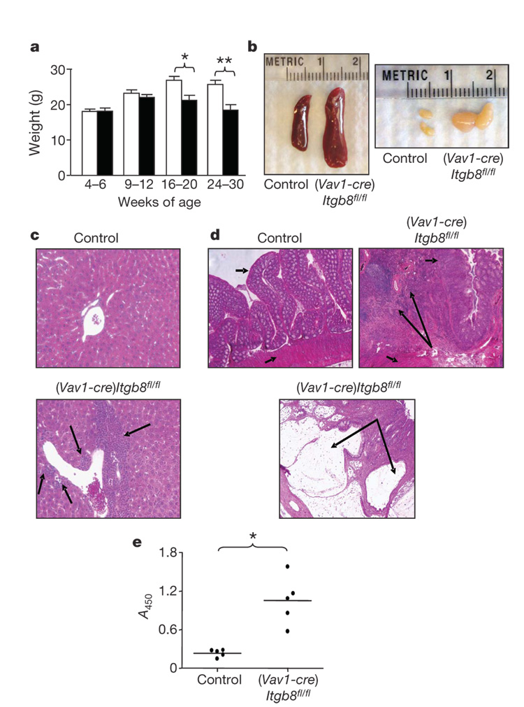
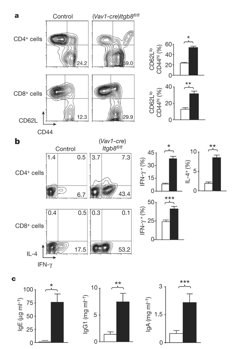
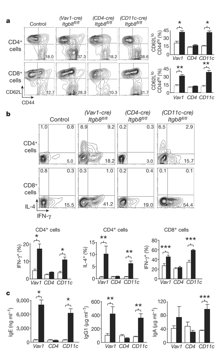
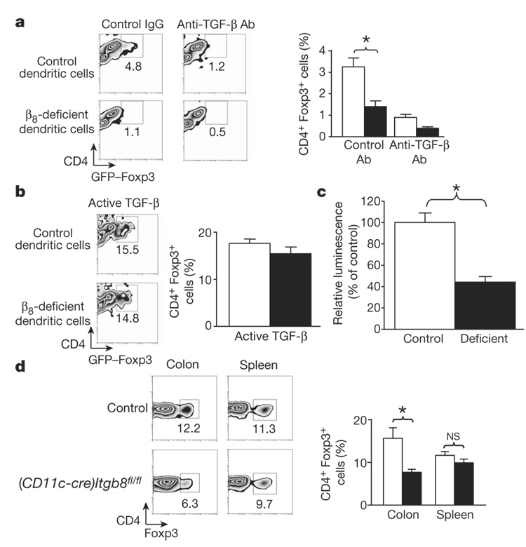
The cytokine transforming growth factor-beta (TGF-beta) is an important negative regulator of adaptive immunity. TGF-beta is secreted by cells as an inactive precursor that must be activated to exert biological effects, but the mechanisms that regulate TGF-beta activation and function in the immune system are poorly understood. Here we show that conditional loss of the TGF-beta-activating integrin alpha(v)beta8 on leukocytes causes severe inflammatory bowel disease and age-related autoimmunity in mice. This autoimmune phenotype is largely due to lack of alpha(v)beta8 on dendritic cells, as mice lacking alpha(v)beta8 principally on dendritic cells develop identical immunological abnormalities as mice lacking alpha(v)beta8 on all leukocytes, whereas mice lacking alpha(v)beta8 on T cells alone are phenotypically normal. We further show that dendritic cells lacking alpha(v)beta8 fail to induce regulatory T cells (T(R) cells) in vitro, an effect that depends on TGF-beta activity. Furthermore, mice lacking alpha(v)beta8 on dendritic cells have reduced proportions of T(R) cells in colonic tissue. These results suggest that alpha(v)beta8-mediated TGF-beta activation by dendritic cells is essential for preventing immune dysfunction that results in inflammatory bowel disease and autoimmunity, effects that are due, at least in part, to the ability of alpha(v)beta8 on dendritic cells to induce and/or maintain tissue T(R) cells.


Figures

Figure 1
Figure 1. (Vav1-cre)Itgb8fl/fl mice develop age-related autoimmunity
a, Weight loss in control and (Vav1-cre)Itgb8fl/fl mice (white, control; black, (Vav1-cre)Itgb8fl/fl; n=7 per group; asterisk, P=0.011; double asterisk, P=0.0026). Error bars represent s.e.m. b, Lymphoid organs of 5-month-old mice. c, Haematoxylin- and eosin-stained sections of livers (10 months, original magnification ×200). Arrows show cellular infiltrates. d, Haematoxylin- and eosin-stained colon sections (9 months, original magnification ×50). Short arrows, epithelium (top arrow) and smooth muscle (bottom arrow); large arrows, cellular infiltrates (top panel) and large cyst (bottom panel). e, ELISA for anti-dsDNA and anti-ribonuclear protein (9–12 months, n=5, P=0.0013).
Figure 2
Figure 2. (Vav1-cre)Itgb8fl/fl mice develop enhanced numbers of activated/memory T cells expressing IL-4 and IFN-γ, and increased serum IgE, IgG1 and IgA levels
a, Activated/memory T cells from spleen (CD62LlowCD44high, 4–6-month-old mice) were analysed by flow cytometry. Representative flow cytometry plots and plotted mean values are shown (white, control; black, (Vav1-cre)Itgb8fl/fl; n=14; asterisk, P=2.7×10⁻¹¹; double asterisk, P=4.9×10⁻⁵). b, IL-4 and IFN-γ levels in T cells from spleen were analysed by intracellular flow cytometry. Representative flow cytometry plots and plotted mean values are shown (white, control; black, (Vav1-cre)Itgb8fl/fl; n=9; asterisk, P=1.8×10⁻⁸; double asterisk, P=1.8×10⁻⁷; triple asterisk, P=0.00048). c, ELISA for IgE, IgG1 and IgA levels in sera (4–6-month-old mice; white, control; black, (Vav1-cre)Itgb8fl/fl; n=4; asterisk, P=0.0028; double asterisk, P=0.011; triple asterisk, P=0.016). All error bars represent s.e.m.
Figure 3
Figure 3. Mice lacking integrin β8 on dendritic cells develop an identical immune phenotype to mice lacking β8integrin on all leukocytes
a, Activated/memory T cells from spleen (CD62LlowCD44high, 4–6-week-old mice) were analysed by flow cytometry. Representative flow cytometry plots and plotted mean values are shown (white, control; black, Itgb8 conditional knockout; n=6 per group; asterisk, P<8.2×10⁻⁴; double asterisk, P<1.4×10⁻⁴). b, IL-4 and IFN-γ levels in T cells from spleen were analysed by intracellular flow cytometry. Representative flow cytometry plots and plotted mean values are shown (white, control; black, Itgb8 conditional knockout; n=6 per group; asterisk, P<0.0063; double asterisk, P=0.0055; triple asterisk, P=0.0045). c, ELISA for IgE, IgG1 and IgA levels in sera (4–6-week-old mice; white, control; black, Itgb8 conditional knockout; n=6; asterisk, P<0.0018; double asterisk, P<0.016; triple asterisk, P=0.0015). All error bars represent s.e.m.
Figure 4
Figure 4. β8-deficient dendritic cells fail to induce TR cells in vitro, and (CD11c-cre)Itgb8fl/fl mice have reduced proportions of TR cells in colonic tissue
a, b, Induction of TR cells (CD4+GFP–Foxp3+) by control or β8-deficient dendritic cells in the presence of control or anti-TGF-β antibody (a) or active TGF- β (b). Representative flow cytometry plots and mean data plots (expressed as percentage of CD4+ cells that expressed GFP–Foxp3) are shown (white, control dendritic cells; black, β8-deficient dendritic cells; n=5; asterisk, P=0.013). c, TGF-β activation by control or β8-deficient dendritic cells, detected using mink lung reporter cells (white, control dendritic cells; black, β8-deficient dendritic cells; n=6; asterisk, P=0.0006). d, TR cell proportions present in spleen or colonic lamina propria. Representative flow cytometry plots and mean data plots (expressed as percentage of CD4+ cells that expressed GFP–Foxp3) are shown (white, control; black, (CD11c-cre)Itgb8fl/fl; n=6; asterisk, P=0.012). NS, not significant. All error bars represent s.e.m.

Comment in

Similar articles

Cited by

References

    1. Shull MM, et al. Targeted disruption of the mouse transforming growth factor-β1 gene results in multifocal inflammatory disease. Nature. 1992;359:693–699. - PMC - PubMed
    1. Marie JC, Liggitt D, Rudensky AY. Cellular mechanisms of fatal early-onset autoimmunity in mice with the T cell-specific targeting of transforming growth factor-β receptor. Immunity. 2006;25:441–454. - PubMed
    1. Li MO, Sanjabi S, Flavell RA. Transforming growth factor-β controls development, homeostasis, and tolerance of T cells by regulatory T cell-dependent and -independent mechanisms. Immunity. 2006;25:455–471. - PubMed
    1. Annes JP, Munger JS, Rifkin DB. Making sense of latent TGFβ activation. J. Cell Sci. 2003;116:217–224. - PubMed
    1. Huang XZ, et al. Inactivation of the integrin β6 subunit gene reveals a role of epithelial integrins in regulating inflammation in the lung and skin. J. Cell Biol. 1996;133:921–928. - PMC - PubMed

Publication types

MeSH terms