. 2007 Sep;37(9):2518-28.
doi: 10.1002/eji.200636892.

Characterisation of the expression and function of the GM-CSF receptor alpha-chain in mice

Affiliations

Characterisation of the expression and function of the GM-CSF receptor alpha-chain in mice

Marcela Rosas et al. Eur J Immunol. 2007 Sep.

Abstract

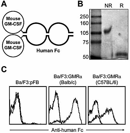
The granulocyte-macrophage colony-stimulating factor (GM-CSF) is a hematopoietic cytokine able to regulate a variety of cell functions including differentiation of macrophages and granulocytes, dendritic cell development and the maintenance of homeostasis. It binds specifically to its receptor, which is composed of a cytokine-specific alpha-chain (GM-CSF receptor alpha-chain, GMRalpha) and a beta-chain shared with the receptors for interleukin-3 and interleukin-5. In this report, we present a comprehensive study of GMRalpha in the mouse. We have found that the mouse GMRalpha is polymorphic and alternatively spliced. In the absence of specific antibodies, we generated a novel chimeric protein containing the Fc fragment of human IgG1 coupled to mouse GM-CSF, which was able to specifically bind to GMRalpha and induce proliferation of GMRalpha-transduced Ba/F3 cells. We used this reagent to perform the first detailed FACS study of the surface expression of mouse GMRalpha by leucocytes. Highest expression was found on monocytes and granulocytes, and variable expression on tissue macrophages. The GM-CSF receptor in mice is specifically expressed by myeloid cells and is useful for the detection of novel uncharacterised myeloid populations. The ability to detect GM-CSF receptor expression in experimental studies should greatly facilitate the analysis of its role in immune pathologies.


Figures

Figure 1
Figure 1
Characterisation of GMRα in the mouse model. (A) Schematic representation of the 11 exons coding for GMRα. The recently published sequence (Ensembl release 40) corresponds to the full-length sequence of C57BL/6 mice whereas Balb/c mice display a polymorphic change in exon 6. We observed two additional splice forms, one with a small deletion and a truncated form, the changes in which are shown schematically. Black arrows indicate coding exons while grey arrows indicate non-coding sequence. (B) Schematic representation of the three predicted isoforms of the mouse GMRα. The full-length isoform is composed of a short cytoplasmic tail, a transmembrane region and an extracellular region, which contains an FN-III domain where there is a polymorphic change at residue 227* (Asp for Balb/c and Gly for C57BL/6). The deletion transcript results in an in-frame deletion in the FN-III domain, but retains a transmembrane domain. The truncated isoform has a premature stop codon.
Figure 2
Figure 2
Functionality of polymorphic forms of GMRα. (A) Full-length GMRα from Balb/c and C57BL/6 mice were retrovirally transduced into Ba/F3 cells. Ba/F3 cells transduced with empty vector or GMRα-encoding vectors were cultured in the presence of 4 ng/mL of GM-CSF (filled circles) or IL-3 (open circles) for the indicated number of days (left panels) or in the indicated concentrations of cytokine for 8 days (right panels). Cell proliferation was measured by determining the percentage of reduction of Alamarblue. (B) The ratio of proliferation in response to GM-CSF and IL-3 was calculated to compare the polymorphic variants; filled squares: Ba/F3:pFB; open circles: Ba/F3:GMRα (Balb/c); filled circles: Ba/F3:GMRα (C57BL/6).
Figure 3
Figure 3
Generation and characterisation of a GM-CSF-Fc fusion protein. (A) Graphic representation of GM-Fc, a chimeric protein generated by fusing the mouse GM-CSF to a mutated Fc region of human IgG1. (B) GM-Fc purity was assessed by Coomassie blue staining of protein resolved under both reducing (R) and non-reducing (NR) conditions by 10% SDS-PAGE. (C) The binding capacity of GM-Fc (grey line) to Ba/F3 cells expressing GMRα from Balb/c or C57BL/6 mice was determined by FACS as indicated previously and compared to the control-Fc protein (black line).
Figure 4
Figure 4
Effect of GM-Fc on Ba/F3 cells expressing full-length GMRα isoforms. (A) Ba/F3:pFB (filled squares), Ba/F3:GMRα (Balb/c; open circles), and Ba/F3:GMRα (C57BL/6; filled circles) were cultured in the presence of either GM-CSF or GM-Fc and cell proliferation was determined using Alamarblue as indicated previously. (B) Expressing percentage Alamarblue reduction obtained when GM-Fc or GM-CSF was used as a growth factor as a ratio shows that both agents have a similar capacity to support proliferation of Ba/F3:GMRα cells. (C) The ability of the C57BL/6 and Balb/c isoforms of GMRα to bind to the cytokine was assessed using GM-Fc and analysed by non-linear regression. Data shown represent pooled data from two independent experiments, and error bars shown the SEM.
Figure 5
Figure 5
Effect of GM-Fc on Ba/F3 cells expressing the deletion and truncation GMRα isoforms. (A) The binding capacity of GM-Fc to Ba/F3 cells expressing the deletion and truncation GMRα from C57BL/6 mice was determined by FACS as indicated previously. (B) Ba/F3:pFB, Ba/F3:GMRα (C57BL/6), the deletion and truncation isoforms (C57BL/6) were cultured in the presence of either GM-CSF, GM-Fc, or IL-3-conditioned medium or in the absence of cytokine. Cell proliferation was determined using Alamarblue as indicated previously.
Figure 6
Figure 6
Detection of GMRα on the surface of blood leukocytes. Cell surface expression of mouse GMRα in different blood cell populations was determined by FACS using GM-Fc or control-Fc. (A) Granulocytes were gated by their SSChighFSCint–high phenotype (left panel 1), and subdivided into neutrophils (Neu, Gr-1highSSChigh; left panel 2), eosinophils (Eos, Gr-1lowSSCv.high; left panel 2) and basophils (Bas, CD49b+IgE+; left panel 3). Binding of control-Fc (middle panels) and GM-Fc (right panels) was then assessed on the three populations as indicated. GM-Fc bound to all three granulocyte populations (B) Monocytes (Mo) were identified by their F4/80+CD11b+ phenotype (left panel) after gating on SSClow cells (not shown). The monocytes were further divided by their expression of Gr-1 and assessed for binding of control-Fc (middle panel) or GM-Fc (right panel). GM-Fc bound well to both major monocyte subsets. (C) GMRα expression was also determined on lymphoid cells. T cells (top panels, gated on CD3+), NK cells (middle panels, gated on CD3CD49b+) and B cells (bottom panels, gated on CD19+) were assessed for binding of GM-Fc (grey lines) and control-Fc (black line). No specific binding of GM-Fc was detected on the lymphocyte populations.
Figure 7
Figure 7
Expression of GMRα by macrophages and characterisation of myeloid cells. (A) Inflammatory peritoneal macrophages were elicited by intraperitoneal administration of thioglycollate broth (top panels) or biogel (middle panels) for 4 days. GMRα expression was analysed by FACS along with the macrophages identified by F4/80 and CD11b profiles (gated regions). Alveolar macrophages were isolated from lung lavage and gated in accordance with the expression of F4/80 and CD11c (bottom panels). GM-Fc (right panels, grey lines), when compared to control-Fc (right panels, black lines), bound specifically and at high levels to the macrophage populations. (B) RAW264.7 cells were stimulated with 100 ng/mL poly(I:C), Pam3CSK4, flagellin or LPS or 5×10⁶ particles/well of zymosan. After 24 h, binding of GM-Fc was assessed by FACS. Data represent the results of four independent experiments and error bars are the SD. Data were analysed by repeated measures one-way ANOVA with Dunnet's post test; *p<0.05, **p<0.01. (C) We used GM-Fc to identify GMRα-expressing cells in the resting peritoneal cavity (upper panels). Gating on GMRα+ cells identified three populations of myeloid cells, which expressed distinct levels of the myeloid markers CD11b and F4/80 (populations 1–3, upper right panel). The physical FSC/SCC profiles of three populations are shown in the lower panels. Population 1 exhibits the classic F4/80highCD11bhigh phenotype of resident peritoneal macrophages. The second F4/80lowCD11bhigh population also expressed MHC class II and exhibited dendritic cell-like characteristics (Dioszeghy et al., manuscript in preparation) and the third rarer population shared the phenotype of eosinophils.

Similar articles

Cited by

References

    1. Hamilton JA, Anderson GP. GM-CSF biology. Growth Factors. 2004;22:225–231. - PubMed
    1. Armitage JO. Emerging applications of recombinant human granulocyte-macrophage colony-stimulating factor. Blood. 1998;92:4491–4508. - PubMed
    1. Trapnell BC, Whitsett JA. GM-CSF regulates pulmonary surfactant homeostasis and alveolar macrophage-mediated innate host defense. Annu. Rev. Physiol. 2002;64:775–802. - PubMed
    1. Reed JAH, Rice WR, Zsengeller ZK, Wert SE, Dranoff G, Whitsett JA. GM-CSF enhances lung growth and causes alveolar type II epithelial cell hyperplasia in transgenic mice. Am. J. Physiol. Lung Cell. Mol. Physiol. 1997;273:L715–L725. - PubMed
    1. Wognum AW, Westerman Y, Visser TP, Wagemaker G. Distribution of receptors for granulocyte-macrophage colony-stimulating factor on immature CD34+ bone marrow cells, differentiating monomyeloid progenitors, and mature blood cell subsets. Blood. 1994;84:764–774. - PubMed

Publication types

MeSH terms

Substances

LinkOut - more resources