Clinical Trial
. 2007 Aug 14;13(30):4141-6.
doi: 10.3748/wjg.v13.i30.4141.

Assessment of T staging and mesorectal fascia status using high-resolution MRI in rectal cancer with rectal distention

Affiliations
Clinical Trial

Assessment of T staging and mesorectal fascia status using high-resolution MRI in rectal cancer with rectal distention

Sheng-Xiang Rao et al. World J Gastroenterol. .

Abstract

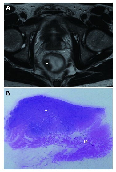
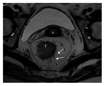
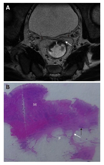
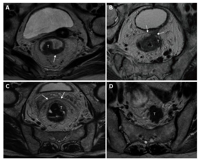
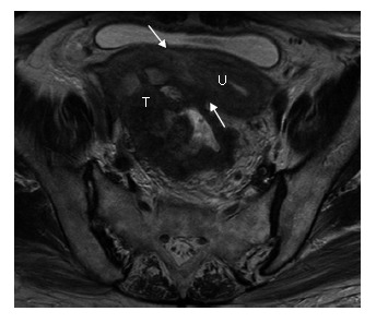
Aim: To determine the accuracy of high-resolution magnetic resonance imaging (MRI) using phased-array coil for preoperative assessment of T staging and mesorectal fascia infiltration in rectal cancer with rectal distention.

Methods: In a prospective study of 67 patients with primary rectal cancer, high-resolution magnetic resonance imaging (in-plane resolution, 0.66 multiply 0.56) with phased-array coil were performed for T-staging and measurement of distance between the tumor and the mesorectal fascia. The assessment of MRI was compared with postoperative histopathologic findings. Sensitivity, specificity, accuracy, positive predictive value, and negative predictive value were evaluated.

Results: The overall magnetic resonance accuracy was 85.1% for T staging and 88% for predicting mesorectal fascia involvement. Magnetic resonance sensitivity, specificity, accuracy, positive predictive value, and negative predictive value was 70%, 97.9%, 89.6%, 93.3% and 88.5% for <or= T2 tumors, 90.5%, 76%, 85.1%, 86.4% and 82.6% for T3 tumors, 100%, 95.2%, 95.5%, 62.5% and 100% for T4 tumors, and 80%, 90.4%, 88%, 70.6% and 94% for predicting mesorectal fascia involvement, respectively.

Conclusion: High-resolution MRI enables accurate preoperative assessment for T staging and mesorectal fascia infiltration in rectal cancer with rectal distention.


Figures

Figure 1
Figure 1
T2-stage rectal cancer in a 58-year-old male patient. A: Axial T2W-TSE MR image (3500/94); B: Photograph of the corresponding histopathologic slice (hematoxylin-eosin stain; original magnification, x 10), showed T2-stage tumor (T) that was confined within the muscularis propria (M).
Figure 2
Figure 2
T2-stage rectal cancer overstaged at MR imaging (3000/98) as a T3-stage tumor in a 70-year-old male patient. MR image depicted tumor (T) with speculations (white arrow) which turned out to desmoplastic reaction without tumor cells at histology.
Figure 3
Figure 3
A: T3-stage rectal cancer without mesorectal fascia involvement in a 64-year-old female patient. Axial T2W-TSE MR image (3000/98) showed tumor in anterior rectal wall (T) with transmural spiculation (black arrow) from tumor into perirectal fat, and the distance to mesorectal fascia (white arrow) is measured ≤2 mm; B: T3-stage rectal cancer with involved mesorectal fascia in a 79-year-old male patient. Axial T2W-TSE MR image (3000/98) showed tumor (T) extending to mesorectal fascia (white arrows); C: T3-stage rectal cancer with involved mesorectal fascia in a 65-year-old male patient. Axial T2W-TSE MR image (3000/98) showed heterogeneous strands are noted in the perirectal fat tissues with the thicken mesorectal fascia; D: T3-stage rectal cancer with extramural deposits in a 62-year-old female patient. Axial T2W-TSE MR image (3000/98) showed extramural deposits (T) in the perirectal space with irregular shape.
Figure 4
Figure 4
A: T3-stage rectal cancer without mesorectal fascia involvement in a 57-year-old female patient. Axial T2W-TSE MR image (3000/98) manifested bulky tumor with broad-based nodular (T) with clear margin (white arrows); B: T3-stage rectal cancer with involved mesorectal fascia in a 79-year-old male patient. Axial T2W-TSE MR image (3000/98) showed tumor (T) extending to mesorectal fascia (white arrows); C: T3-stage rectal cancer with involved mesorectal fascia in a 65-year-old male patient. Axial T2W-TSE MR image (3000/98) showed heterogeneous strands are noted in the perirectal fat tissues with the thicken mesorectal fascia; D: T3-stage rectal cancer with extramural deposits in a 62-year-old female patient. Axial T2W-TSE MR image (3000/98) showed extramural deposits (T) in the perirectal space with irregular shape.
Figure 5
Figure 5
T4-stage rectal cancer with fixation to uterus in a 76-year-old female patient. Axial T2W-TSE MR image (3000/98) showed direct invasion (white arrows) of tumour (T) into uterus (U).

References

    1. Li M, Gu J. Changing patterns of colorectal cancer in China over a period of 20 years. World J Gastroenterol. 2005;11:4685–4688. - PMC - PubMed
    1. Adam IJ, Mohamdee MO, Martin IG, Scott N, Finan PJ, Johnston D, Dixon MF, Quirke P. Role of circumferential margin involvement in the local recurrence of rectal cancer. Lancet. 1994;344:707–711. - PubMed
    1. Nagtegaal ID, Marijnen CA, Kranenbarg EK, van de Velde CJ, van Krieken JH. Circumferential margin involvement is still an important predictor of local recurrence in rectal carcinoma: not one millimeter but two millimeters is the limit. Am J Surg Pathol. 2002;26:350–357. - PubMed
    1. Compton CC, Greene FL. The staging of colorectal cancer: 2004 and beyond. CA Cancer J Clin. 2004;54:295–308. - PubMed
    1. Heald RJ, Ryall RD. Recurrence and survival after total mesorectal excision for rectal cancer. Lancet. 1986;1:1479–1482. - PubMed

Publication types