. 2007 Aug;4(8):e253.
doi: 10.1371/journal.pmed.0040253.

Expansion of regulatory T cells in patients with Langerhans cell histiocytosis

Affiliations

Expansion of regulatory T cells in patients with Langerhans cell histiocytosis

Brigitte Senechal et al. PLoS Med. 2007 Aug.

Abstract

Background: Langerhans cell histiocytosis (LCH) is a rare clonal granulomatous disease that affects mainly children. LCH can involve various tissues such as bone, skin, lung, bone marrow, lymph nodes, and the central nervous system, and is frequently responsible for functional sequelae. The pathophysiology of LCH is unclear, but the uncontrolled proliferation of Langerhans cells (LCs) is believed to be the primary event in the formation of granulomas. The present study was designed to further investigate the nature of proliferating cells and the immune mechanisms involved in the LCH granulomas.

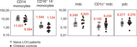
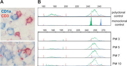
Methods and findings: Biopsies (n = 24) and/or blood samples (n = 25) from 40 patients aged 0.25 to 13 y (mean 7.8 y), were studied to identify cells that proliferate in blood and granulomas. We found that the proliferating index of LCs was low ( approximately 1.9%), and we did not observe expansion of a monocyte or dendritic cell compartment in patients. We found that LCH lesions were a site of active inflammation, tissue remodeling, and neo-angiogenesis, and the majority of proliferating cells were endothelial cells, fibroblasts, and polyclonal T lymphocytes. Within granulomas, interleukin 10 was abundant, LCs expressed the TNF receptor family member RANK, and CD4(+) CD25(high) FoxP3(high) regulatory T cells (T-regs) represented 20% of T cells, and were found in close contact with LCs. FoxP3(+) T-regs were also expanded compared to controls, in the blood of LCH patients with active disease, among whom seven out of seven tested exhibited an impaired skin delayed-type hypersensitivity response. In contrast, the number of blood T-regs were normal after remission of LCH.

Conclusions: These findings indicate that LC accumulation in LCH results from survival rather than uncontrolled proliferation, and is associated with the expansion of T-regs. These data suggest that LCs may be involved in the expansion of T-regs in vivo, resulting in the failure of the host immune system to eliminate LCH cells. Thus T-regs could be a therapeutic target in LCH.


Conflict of interest statement

Competing Interests: The authors have declared that no competing interests exist.

Figures

Figure 1
Figure 1. Proliferating Cells in LCH Granuloma are Mostly Endothelial Cells, Fibroblasts, and T Cells
Paraffin-embedded and frozen sections were stained with antibodies against Ki-67 (which label proliferating cells), CD1a (LCs), CD3 (T cells), CD20 (B cells), CD68, CD31, and CD34 (endothelial cells). (A) Double immunostaining of paraffin-embedded section from LCH eosinophilic granulomas with anti-Ki-67 Ab, (brown nuclear staining) and with anti-CD1a Ab (upper images, blue staining) or anti-CD3 Ab (lower images, blue staining). Open arrowheads indicate double-stained cells, black arrowheads indicate Ki-67+ cells with an endothelial morphology. (B) Histogram represents percentage of CD1a+ cells and of CD3+ cells labeled with Ki-67 (n = 15). (C) Histogram represents percentage of proliferating cells (Ki-67+) that express CD1a, CD3, CD20, or CD68 (n = 15). (D) Histogram represents percentage of proliferating cells (Ki-67+) that are endothelial cells, interstitial cells (fibroblasts), and other types based on morphological examination. (E) Immunolabeling of blood vessels on paraffin-embedded section from LCH eosinophilic granulomas with CD34 (left) and CD31 (right) antibodies. (F) Proliferating Ki-67+ cells (brown nuclear staining) with a fibroblast-cell morphology in an eosinophilic granuloma.
Figure 2
Figure 2. Putative LC Precursors are Detected at Normal Frequencies in the Peripheral Blood of Naïve LCH Patients
CD14+ monocytes (DR+, CD14+, CD16lo) and CD16+ monocytes (DR+, CD14, CD16hi), MDCs (DR+, CD11c+, lin), CD1c+ MDCs (DR+, CD1c+, CD11c+, lin), and PDCs (DR+, BDCA-2+, CD123+) from naïve (untreated) LCH patients and age-matched control children were studied by five-color flow cytometry. Data are represented as the percentage of PBMCs. p-Values (Kruskal-Wallis test) for the comparison between naïve (untreated) LCH patients and age-matched control children were not significant either in the number or in the frequency of DC and monocyte subsets present in the peripheral blood.
Figure 3
Figure 3. Polyclonal T Cells Infiltrate LCH Granuloma in Close Contact with LCs
(A) Double immunohistochemical labeling of frozen sections from an eosinophilic granuloma with anti-CD3 (brown) and anti-CD1a antibodies (blue). (B) T cell receptor gamma rearrangement was determined on the DNA extracted from frozen biopsies from four patients. Fluorescent profiles for Vgfl/Vg10 PCR using fluorescent Jg primers (JgP, red; Jg1/2, green; JgP1/2, blue) are shown; all the biopsies display a polyclonal profile in comparison to polyclonal and monoclonal positive controls.
Figure 4
Figure 4. CD4+ CD25+ FoxP3high T Cells Accumulate in LCH Granulomas
(A) Identification of CD4+ CD25+ FoxP3+ T cells by flow cytometry on fresh granuloma tissue. Dot plots are representative of similar data obtained in three patients (11, 12, and 29). (B) Histograms represent percentage of FoxP3+ T cells among CD3+ T cells in eosinophilic granuloma: 17.2% ± 3.36% of T lymphocytes expressed FoxP3 (n = 6). (C) Left image, immunohistochemical staining on a paraffin-embedded section with FoxP3 antibodies showing FoxP3+ T cell (arrow, brown nucleus) in close contact with histiocytes (arrowhead). Right images, double immunofluorescence labelling with anti-FoxP3 (green nucleus) and anti-CD3+, anti-CD25+, or anti-CD1a (red) in frozen sections. (D) Real-time PCR assay showing a high level of IL10 transcripts in eosinophilic granulomas (n = 12 patients). (E) T cells were isolated by CD3+ selection from a fresh LCH biopsy (patient 12; LCH T cells) and from the peripheral blood of a healthy blood donor (Donor T cells) and were stimulated by soluble OKT3 (Anti-CD3) in the presence of CD14+ monocytes. Unsorted T cells from an LCH granuloma (LCH T cells) caused an approximate 50% decrease in the proliferation of normal T cells. Filled bar, healthy donor T cells alone, open bar LCH T cells alone, hatched bar, mix of healthy donor T cells and LCH T cells. (F) Immunohistochemistry on paraffin-embedded section from an eosinophilic granuloma using RANK (upper) and RANKL antibodies (bottom) (n = 9 patients). (G) Double immunolabeling with Ki-67 and FoxP3 antibodies of frozen sections from an eosinophilic granuloma (n = 2 patients).
Figure 5
Figure 5. Regulatory CD4+ CD25+ FoxP3+ T Cells Are Expanded in the Peripheral Blood of LCH Patients
(A and B) Flow cytometry analysis of CD4+/CD25hi FoxP3high cells in the blood of LCH patients in comparison to healthy controls. (A) Representative dot plots from the blood of one patient and one control. (B) CD3 and CD4 lymphocyte counts were similar in naïve (untreated) LCH patients at diagnosis and in control children, but the absolute number and the frequency of T-regs were significantly higher in the blood of naïve LCH patients at diagnosis than in patients with LCH under treatment, in control children, and in children with SHML. *Chi-squared = 7.125 with 1 df, p = 0.0076; **Chi-squared = 13.714 with 1 df, p < 0.001; ***Chi-squared = 6.682 with 1 df, p = 0.0097. (C) Regulatory activity of CD3+ CD4+ CD25hi T cells isolated from the blood of LCH patients. Proliferation of control T cells (Donor T cells) induced by soluble OKT3 (Anti CD3) in the presence of CD14+ monocytes was inhibited by the addition of sorted CD3+ CD4+ CD25hi T cells (LCH CD4/CD25hi) isolated by positive selection from the blood of a patient with LCH, but not by CD3+ CD4+ CD25low T cells (LCH CD4/25lo) isolated from the blood of the same patient. *p < 0.02, **p = 0.01.

References

    1. The French Langerhans' Cell Histiocytosis Study Group. A multicentre retrospective survey of Langerhans' cell histiocytosis: 348 cases observed between 1983 and 1993. Arch Dis Child. 1996;75:17–24. - PMC - PubMed
    1. Lichtenstein L. Histiocytosis X: Integration of eosinophilic granuloma of bone, Letterer-Siwe disease, and Schuller-Christian disease as related manifestations of a single nosologic entity. AMA Arch Pathol. 1953;56:84–102. - PubMed
    1. Favara BE, Steele A. Langerhans cell histiocytosis of lymph nodes: A morphological assessment of 43 biopsies. Pediatr Pathol Lab Med. 1997;17:769–787. - PubMed
    1. Geissmann F, Thomas C, Emile JF, Micheau M, Canioni D, et al. Digestive tract involvement in Langerhans cell histiocytosis. The French Langerhans Cell Histiocytosis Study Group. J Pediatr. 1996;129:836–845. - PubMed
    1. Geissmann F, Lepelletier Y, Fraitag S, Valladeau J, Bodemer C, et al. Differentiation of Langerhans cells in Langerhans cell histiocytosis. Blood. 2001;97:1241–1248. - PubMed

Publication types