. 2008 Jan;10(1):177-87.
doi: 10.1111/j.1462-5822.2007.01028.x. Epub 2007 Aug 14.

The bundlin pilin protein of enteropathogenic Escherichia coli is an N-acetyllactosamine-specific lectin

Affiliations

The bundlin pilin protein of enteropathogenic Escherichia coli is an N-acetyllactosamine-specific lectin

Romney M Hyland et al. Cell Microbiol. 2008 Jan.

Abstract

Synthetic N-acetyllactosamine (LacNAc) glycoside sequences coupled to BSA competitively inhibit enteropathogenic Escherichia coli (EPEC) localized adherence (LA) to human intestinal biopsy specimens and tissue culture cell monolayers. The LacNAc-specific adhesin appears to be associated with the bundle-forming pili (BFP) expressed by EPEC during the early stages of colonization. Herein, we report that recombinant bundlin inhibits EPEC LA to HEp-2 cells and binds to HEp-2 cells. Recombinant bundlin also binds, with millimolar association constants (K(assoc)), to synthetic LacNAc-Benzene and LacNAc-O(CH(2))(8)CONH(2) glycosides as assessed in the gas phase by nanoelectrospray ionization mass spectrometry. Furthermore, LacNAc-BSA inhibits LA only of EPEC strains that express alpha bundlin alleles, suggesting putative locations for the LacNAc-binding pocket in the alpha bundlin monomer. Collectively, these results suggest that alpha bundlin possesses lectin-like properties that are responsible for LacNAc-specific initial adherence of alpha bundlin-expressing EPEC strains to host intestinal epithelial cells.


Figures

Fig. 1
Fig. 1
EPEC (strain E2348/69) LA to HEp-2 cells in the presence of 280 μM LacNAc-BSA (white bars) or unconjugated BSA (black bars). LA assays were performed by three protocols: (1) 45 min incubation on HEp-2 cells in the presence or absence of glycoconjugate, (2) 3 h incubation on HEp-2 cells in the presence or absence of glycoconjugate, and (3) 3 h incubation on HEp-2 cells in the presence or absence of 280 μM glycoconjugate with additional 280 μM glycoconjugate added after 2 h. For each experiment, 100 HEp-2 cells were observed in triplicate for LA EPEC, defined as microcolonies composed of at least four bacteria. Error bars represent the standard deviations from the means.
Fig. 2
Fig. 2
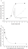
Radio-receptor binding isotherm (A) and competition binding assays (B) using recombinant bundlin. A. Increasing amounts of radio-iodinated bundlin were applied to HEp-2 cell monolayers in the presence and absence of 500-fold excess unlabelled bundlin. Each of the data points represents specifically bound cpm, i.e. the difference between the cpm bound at each concentration in the absence of unlabelled bundlin minus the residual cpm bound in the presence of unlabelled bundlin. Data from the isotherm were converted into a linear (r2 = 0.9644) Hill plot, shown in the inset. B. Radio-iodinated bundlin (3.4 nM) was added to HEp-2 cell monolayers in the presence of increasing concentrations of unlabelled bundlin. In both panels, each data point represents the average of triplicate determinations and the error bars indicate the standard error of the individual means.
Fig. 3
Fig. 3
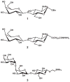
Structures of LacNAc-Bn (1), LacNAc-O(CH2)8CONH2 (2) and the αGal(1,4)βGal(1,4)βGlc-SiMe3 (Pk blood group trisaccharide) (3).
Fig. 4
Fig. 4
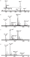
NanoES mass spectra of aqueous solutions of 20 μM bundlin in the absence (A) or presence of 115 μM 1 (B), 130 μM 2 (C) or 150 μM 3 (D).
Fig. 5
Fig. 5
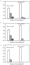
Distribution of ligand 1 (A), 2 (B) and 3 (C) bound to bundlin and Pref, determined directly from the mass spectra. Also shown are the distributions of ligands bound to bundlin after correction for non-specific ligand binding based on the interaction between Pref and the ligands.
Fig. 6
Fig. 6
A. LA to HEp-2 cells of EPEC strains harbouring different bfpA alleles. BFP expression was induced by pre-incubating the EPEC strains in DMEM for 30 min in the CO2 incubator. Subsequently, EPEC cells were added to subconfluent HEp-2 cell monolayers and incubated for 45 min. LA was scored as described previously (Vanmaele et al., 1999), and the data are presented as the percentage of HEp-2 cells harbouring LA EPEC. B. The effect of 280 μM LacNAc-BSA on EPEC LA. Subsequent to the induction of BFP expression, EPEC were incubated with either 280 μM LacNAc-BSA or BSA for 30 min and then applied to HEp-2 cell monolayers. Data were normalized to those obtained when underivatized BSA was used instead of LacNAc-BSA. Each experiment was performed in triplicate, and the error bars represent the standard deviations from the means.
Fig. 7
Fig. 7
BFP expression by strain E2348/69 (A), UMD901 (B) and UMD901 complemented in trans with pRPA100 (C), pXLW13 (D), pXLW16 (E) and pXLW17 (F). Scale bars represent 200 nm.
Fig. 8
Fig. 8
Bundlin expression by strain E2348/69 (A), UMD901 (B), UMD901 (pRPA100) (C), UMD901 (pXLW13) (D), UMD901 (pXLW16) (E) and UMD901 (pXLW17) (F) as detected by Western immunoblot analysis using polyclonal rabbit antisera prepared using bundlin from EPEC strain E2348/69. Sample loading on the gels was normalized using maltose-binding protein (MBP) as an internal standard as described previously (Hyland et al., 2006a). Blots are representative of three experiments.
Fig. 9
Fig. 9
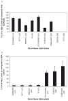
LA of strain E2348/69, UMD901, UMD901 (pRPA100), UMD901 (pXLW13), UMD901 (pXLW16) and UMD901 (pXLW17) in the presence of 280 μM LacNAc-BSA (black bars) and BSA (white bars) in the 3 h LA assay, and in the presence of 280 μM LacNac-BSA (grey bars) or BSA (hatched bars) in the 45 min LA assay. Each experiment was performed in triplicate, and the error bars represent the standard deviations from the means. The reduction in LA in the presence of LacNAc-BSA is significant in the E2348/69 and UMD901 (pRPA100) in the 45 min experiments (P < 0.001, Student’s t-test). Strains UMD901 and UMD901 (pXLW13) did not display LA to HEp-2 cells after 3 h, but rather bound to the cells as individual bacteria, and data in these cases are given as percentage HEp-2 cells with adherent bacteria.

Similar articles

Cited by

References

    1. Anantha RP, Stone KD, Donnenberg MS. Effects of bfp mutations on biogenesis of functional enteropathogenic Escherichia coli type IV pili. J Bacteriol. 2000;182:2498–2506. - PMC - PubMed
    1. Armstrong GD, Peppler MS. Maintenance of biological activity of pertussis toxin radioiodinated while bound to fetuin-agarose. Infect Immun. 1987;55:1294–1299. - PMC - PubMed
    1. Armstrong GD, Mulvey GL, Marcato P, Griener TP, Kahan MC, Tennent GA, et al. Human serum amyloid P component protects against Escherichia coli O157:H7 Shiga toxin 2 in vivo: therapeutic implications for hemolytic-uremic syndrome. J Infect Dis. 2006;193:1120–1124. - PubMed
    1. Barnett Foster D, Philpott D, Abul-Milh M, Huesca M, Sherman PM, Lingwood CA. Phosphatidylethanolamine recognition promotes enteropathogenic E. coli and enterohemorrhagic E. coli host cell attachment. Microb Pathog. 1999;27:289–301. - PubMed
    1. Bieber D, Ramer SW, Wu CY, Murray WJ, Tobe T, Fernandez R, Schoolnik GK. Type IV pili, transient bacterial aggregates, and virulence of enteropathogenic Escherichia coli. Science. 1998;280:2114–2118. - PubMed

Publication types

MeSH terms

LinkOut - more resources