. 2007 Aug 15:8:82.
doi: 10.1186/1471-2474-8-82.

Management of insertional Achilles tendinopathy through a Cincinnati incision

Affiliations

Management of insertional Achilles tendinopathy through a Cincinnati incision

Michael R Carmont et al. BMC Musculoskelet Disord. .

Abstract

Background: About 10% of patients not responding to 3-6 months of conservative management for insertional Achilles tendinopathy undergo surgery. Traditionally, surgery of the Achilles tendon is performed through longitudinal extensile incisions. Such surgery is prone to the complications of wound healing, wound breakdown and iatrogenic nerve injury.

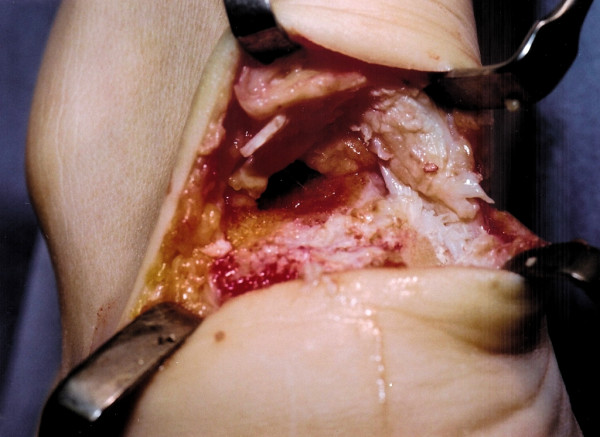
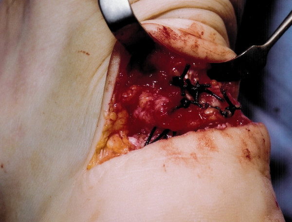
Methods: We describe our current method of exposure of the Achilles tendon insertion and debridement of the peritendinous and tendon tissue with osteotomy of the calcaneum through a transverse skin incision at the level of the Achilles insertion.

Results: This method has been used since 2002 on over 40 patients for exposure of the Achilles tendon insertion and the distal Achilles tendon.

Conclusion: The Cincinnati incision allows adequate exposure, has minimal risk of symptomatic iatrogenic nerve injury, and has minimal problems related to the scar.


Figures

Figure 1
Figure 1
A 5 cm semi-circumferential incision is made at the level of the Achilles tendon insertion over the tendinopathic area.
Figure 2
Figure 2
After dissection and debridement, the tendinopathic tendon insertion (solid arrow) is detached from the calcaneum.
Figure 3
Figure 3
The prominent calcaneal tuberosity is detached from the calcaneum using an osteotome.
Figure 4
Figure 4
The underlying cancellous bone is now exposed.
Figure 5
Figure 5
A 2.5 mm drill is used to pass holes (solid arrows) through the calcaneum to allow suture fixation to the calcaneum. The calcaneum is devoid of soft tissue as all tendinopathic tissue has been debrided.
Figure 6
Figure 6
The exposed surface of the calcaneum is smoothed using a wrasp.
Figure 7
Figure 7
Strong absorbable suture material is used to reinsert the Achilles tendon to the calcaneum.
Figure 8
Figure 8
The wound is closed using subcuticular sutures.
Figure 9
Figure 9
Several months after surgery the mature transverse scar is barely visible.

References

    1. Clain MR, Baxter DE. Achilles tendonitis. Foot Ankle. 1992;13:482–487. - PubMed
    1. Kvist , Lehto , Jozsa , et al. Chronic Achilles paratendionsis: an immuno histologic study of fibronectin and fibrinogen. Am J Sport Med. 1988;16:616–623. doi: 10.1177/036354658801600611. - DOI - PubMed
    1. Maffulli N, Testa V, Capasso G, Sullo A. Calcific insertional Achilles tendinopathy. Am J Sports Med. 2004;32:174–181. doi: 10.1177/0363546503258923. - DOI - PubMed
    1. Fahlstrom M, Jonsson P, Lorentzon R, Alfredson H. Cronic Achilles pain treated with eccentric calf muscle training. Knee Sports Surg Traumatol Arthrosc. 2003;11:327–333. doi: 10.1007/s00167-003-0418-z. - DOI - PubMed
    1. Furia JP. High energy extracorporal shock wave therapy as a treatment for insertional Achilles tendinopathy. Am J Sports Med. 2006;34:733–740. doi: 10.1177/0363546505281810. - DOI - PubMed