. 2007 Aug 15:7:48.
doi: 10.1186/1472-6750-7-48.

One-step selection of Vaccinia virus-binding DNA aptamers by MonoLEX

Affiliations

One-step selection of Vaccinia virus-binding DNA aptamers by MonoLEX

Andreas Nitsche et al. BMC Biotechnol. .

Abstract

Background: As a new class of therapeutic and diagnostic reagents, more than fifteen years ago RNA and DNA aptamers were identified as binding molecules to numerous small compounds, proteins and rarely even to complete pathogen particles. Most aptamers were isolated from complex libraries of synthetic nucleic acids by a process termed SELEX based on several selection and amplification steps. Here we report the application of a new one-step selection method (MonoLEX) to acquire high-affinity DNA aptamers binding Vaccinia virus used as a model organism for complex target structures.

Results: The selection against complete Vaccinia virus particles resulted in a 64-base DNA aptamer specifically binding to orthopoxviruses as validated by dot blot analysis, Surface Plasmon Resonance, Fluorescence Correlation Spectroscopy and real-time PCR, following an aptamer blotting assay. The same oligonucleotide showed the ability to inhibit in vitro infection of Vaccinia virus and other orthopoxviruses in a concentration-dependent manner.

Conclusion: The MonoLEX method is a straightforward procedure as demonstrated here for the identification of a high-affinity DNA aptamer binding Vaccinia virus. MonoLEX comprises a single affinity chromatography step, followed by subsequent physical segmentation of the affinity resin and a single final PCR amplification step of bound aptamers. Therefore, this procedure improves the selection of high affinity aptamers by reducing the competition between aptamers of different affinities during the PCR step, indicating an advantage for the single-round MonoLEX method.


Figures
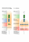
Figure 1
Figure 1
Schematic presentation of MonoLEX in comparison to SELEX. Based on a combinatory oligonucleotide library, SELEX comprises several cycles of target binding, elution and amplification of putative aptamers. In contrast, MonoLEX starts with one affinity chromatography to sort non-binding oligonucleotides, low-affinity aptamers and high-affinity aptamers. Highly affine aptamers are amplified once and characterized further by an aptamer blot assay.
Figure 2
Figure 2
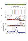
Identification of high-affinity-binding aptamers by MonoLEX. A combinatorial DNA library (64 b in length) was applied to an affinity capillary column coated with complete heat-inactivated VACV. Bound aptamers were desorbed from cut column slices and amplified by real-time PCR. (a) Data derived from different segments along the affinity column show a cumulation of aptamers in distinct segments while other segments do not amplify bound aptamers. Color labels indicate segments which were used for further evaluation of the aptamer pools. Figure 2b to 2d show the change of fluorescence per change of temperature plotted versus the temperature. (b) Melting temperature analysis of polyclonal aptamer pools after PCR amplification showing aptamer pools with different nucleic acid composition. In two of the pools (A38 and A77) more than one melting temperature maximum was observed, indicating different aptamer sub-pools. (c) Melting temperature analysis after aptamer dot blotting with the target molecule and repeated amplification. In A38 and A77 only one of the two sub-pools was amplified, indicating that one aptamer is more efficiently amplified after binding. The inset shows the preceding amplification results of an aptamer blotting assay with VACV (red solid line) and a negative control (black dotted line). The relative fluorescence is plotted vs. the cycle number. (d) Melting profile of the chemically synthesized and further characterized A38 with its predicted two-dimensional structure.
Figure 3
Figure 3
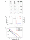
In vitro-binding studies of A38. (a) Dot blot of VACV (VR-1536), non-infective cell culture supernatant (CC) and CMV, followed by incubation of biotin-conjugated A38 in various concentrations and labeling with a streptavidin peroxidase conjugate. (b) Surface Plasmon Resonance measurements (Biacore) of A38. Sequence of injections (injection start): A38 (150 s); VACV (1050 s); 50 mM sodium carbonate (2320 s); 1:10 diluted VACV (4080 s). The overlaid VACV net binding curves are shown on the right hand side. (c) Fluorescence Correlation Spectroscopy (FCS). Normalized autocorrelation functions (ACF) of tetramethylrhodamine isothiocyanate (TMR): free in solution (black), coupled to A38 alone (blue), coupled to A38 with added CMV (brown), coupled to A38 with added VACV (red), and directly coupled to VACV (green). Control experiments with CMV (brown) and 200-nm latex beads (not shown) mixed with TMR-conjugated A38 showed identical diffusion times to those for TMR-labeled A38 alone, indicating a binding exclusively to VACV particles.
Figure 4
Figure 4
In vitro antiviral activity of A38. Immunofluorescence microscopy of VACV-infected Hep2 cells incubated with A38 after 24 hours. (a) Cells infected with VACV at MOI of 0.1, (b) cells incubated with a mixture of VACV and 2.5 μM A38 or (c) VACV and 2.5 μM of the complete template library. (d) Control of non-infected cells. Bar, 100 μm. Red cells are counterstained with Evans's Blue.
Figure 5
Figure 5
Antiviral activity of A38 against various OPV. OPV were propagated in cell culture in presence or absence of A38 as described above. The degree of infection in comparison to a positive control in the absence of A38 was determined by enumerating the number of infected cells (IFA, black bars), quantification of intracellular poxvirus DNA by real-time PCR (red bars) or determination of the poxvirus titer in the corresponding supernatant (green bars).
Figure 6
Figure 6
Stability of DNA aptamer A38. A38 was incubated for 24 h at 37°C in water or, in comparison, in serum. No significant shift of CT value was observed by quantitative real-time PCR for the serum-incubated aptamer (brown lines, n = 3) in comparison to the fresh aptamer A38 (blue lines, n = 2).

Similar articles

Cited by

References

    1. Jahrling PB, Fritz EA, Hensley LE. Countermeasures to the bioterrorist threat of smallpox. Curr Mol Med. 2005;5:817–826. - PubMed
    1. Yang G, Pevear DC, Davies MH, Collett MS, Bailey T, Rippen S, Barone L, Burns C, Rhodes G, Tohan S, Huggins JW, Baker RO, Buller RL, Touchette E, Waller K, Schriewer J, Neyts J, DeClercq E, Jones K, Hruby D, Jordan R. An orally bioavailable antipoxvirus compound (ST-246) inhibits extracellular virus formation and protects mice from lethal orthopoxvirus Challenge. J Virol. 2005;79:13139–13149. - PMC - PubMed
    1. Zhang Z, Blank M, Schluesener HJ. Nucleic acid aptamers in human viral disease. Arch Immunol Ther Exp (Warsz ) 2004;52:307–315. - PubMed
    1. Breaker RR. Natural and engineered nucleic acids as tools to explore biology. Nature. 2004;432:838–845. - PubMed
    1. Hesselberth JR, Miller D, Robertus J, Ellington AD. In vitro selection of RNA molecules that inhibit the activity of ricin A-chain. J Biol Chem. 2000;275:4937–4942. - PubMed

Publication types

MeSH terms