Comparative Study
. 2007 Aug;15(8):928-41.
doi: 10.1016/j.str.2007.06.010.

NikD, an unusual amino acid oxidase essential for nikkomycin biosynthesis: structures of closed and open forms at 1.15 and 1.90 A resolution

Affiliations
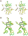
Comparative Study

NikD, an unusual amino acid oxidase essential for nikkomycin biosynthesis: structures of closed and open forms at 1.15 and 1.90 A resolution

Christopher J Carrell et al. Structure. 2007 Aug.

Abstract

NikD is an unusual amino-acid-oxidizing enzyme that contains covalently bound FAD, catalyzes a 4-electron oxidation of piperideine-2-carboxylic acid to picolinate, and plays a critical role in the biosynthesis of nikkomycin antibiotics. Crystal structures of closed and open forms of nikD, a two-domain enzyme, have been determined to resolutions of 1.15 and 1.9 A, respectively. The two forms differ by an 11 degrees rotation of the catalytic domain with respect to the FAD-binding domain. The active site is inaccessible to solvent in the closed form; an endogenous ligand, believed to be picolinate, is bound close to and parallel with the flavin ring, an orientation compatible with redox catalysis. The active site is solvent accessible in the open form, but the picolinate ligand is approximately perpendicular to the flavin ring and a tryptophan is stacked above the flavin ring. NikD also contains a mobile cation binding loop.


Figures

Figure 1
Figure 1
Structure of nikkomycin and streptogramin antibiotics (top panel) and the nikD-catalyzed oxidation of P2C to picolinate (bottom panel). The peptidyl portion of each antibiotic is shown in red with a black box indicating atoms derived from L-lysine. The variable nikkomycin nucleoside moiety is shown in blue. The DHP intermediate shown in the bottom panel is one of 6 possible isomers.
Figure 2
Figure 2
Stereo views of the electron density of closed and open forms of nikD. The electron density surrounding the flavin ring of FAD, its covalent attachment to Cys321 and the endogenous ligand modeled as picolinate is shown. The atoms are colored with nitrogen blue, oxygen red, sulfur yellow and carbon green. (A) Refined |2Fo|-|Fc| electron density at 1.15 Å resolution of the closed form of nikD, contoured at 1 σ (cyan) and 3 σ (red). (B) Refined |2Fo|-|Fc| electron density at 1.90 Å resolution of the open form of nikD, contoured at 1 σ (cyan).
Figure 3
Figure 3
Closed form of nikD. (A) Stereo ribbon drawing with the flavin binding domain colored magenta and the catalytic domain colored cyan. The FAD and picolinate ligand are also included as stick figures with the same coloring scheme as in Fig. 2. (B) Closeup stereo view of the active site in the closed form of nikD. The putative picolinate ligand sits above the flavin ring in a pocket defined by the aromatic side chains of Trp355, Phe242, Phe244, Tyr258, Phe260. Above picolinate and the aromatic cluster are the side chains of Asn100 and Asp276. Oxygen and nitrogen atoms are colored red and blue, respectively; for FAD and picolinate, carbon atoms are colored green and for the side chains they are colored yellow. Hydrogen bonds are represented by dashed lines. (C) Close-up stereo view of the active site of MSOX containing the inhibitor pyrrole-2-carboxilic acid (PCA) (pdb code 1eli), oriented in a manner similar to that of the closed form of nikD, as shown in Fig. 3B. Sulfur is yellow and all other atoms are colored using the same scheme as in Fig. 3B. Side chains that surround PCA and correspond approximately in position to the aromatic side chain cluster Trp355, Phe242, Phe244, Tyr258, Phe260 found in nikD are included. Hydrogen bonds are represented as black dashed lines.
Figure 4
Figure 4
Comparison of the open and closed forms of nikD. (A) Cα trace of the open form of nikD (blue) superimposed on that of the closed form (magenta). Included are stick figures of FAD and picolinate for the closed form only, colored as in Fig. 2. Carbon, nitrogen, oxygen and phosphorus atoms are colored green, blue, red and gold, respectively. (B) Surface representation of the closed form of nikD. There is a small channel visible that leads into the active site. (C) Open form surface representation of nikD. The indole ring of Trp355 (shown as a stick figure) is exposed to the solvent.
Figure 5
Figure 5
Active sites of open and closed forms of nikD. (A) Close-up view of the active site in the open form of nikD. The chemical groups shown and the atom coloring scheme used are the same as in Fig. 3B, except for the addition of two water molecules (red spheres). Hydrogen bonds are represented by dashed lines. (B) Superimposition of the active sites in the open and closed forms of nikD. Carbon atoms are colored green and yellow in the open and closed forms, respectively. In each form, the same color is used for oxygen (red) or nitrogen (blue).
Figure 6
Figure 6
Configuration of the 259-271 segment in the sodium-containing wild-type and sodium-free SeMet closed forms of nikD. (A) Wild-type closed nikD with a sodium ion (violet) coordinated by carbonyl oxygen atoms of Ala266 and Gly268 plus 4 water molecules, and His262 forming a salt bridge with Glu269. Nitrogen, oxygen and carbon atoms are colored blue, red and yellow, respectively. Sodium coordination and hydrogen bonds are shown as black dashed lines. The non-bonding distance from Phe260 to the flavin N(5) atom is shown as a red dashed line. (B) SeMet nikD without bound sodium in which the His262 side chain replaces the sodium ion and forms hydrogen bonds to the carbonyl oxygens of Ala266, Gly268 and Phe270. The atom coloring is the same as in part A.
Figure 7
Figure 7
The reaction scheme shows the mechanism postulated for conversion of P2C to picolinate, as discussed in the text. The inset in the bottom right-hand corner shows the positions of FAD and picolinate, in the same orientation as shown in Fig. 3B. Distances (Å) between N(5) of FAD and the indicated atoms in picolinate are shown in magenta. Angles (°) defined by N(5) and N(10) in FAD and the indicated atoms in picolinate are shown in cyan.

References

    1. Barriere JC, Berthaud N, Beyer D, Dutka-Malen S, Paris JM, Desnottes JF. Recent developments in streptogramin research. Curr Pharm Des. 1998;4:155–180. - PubMed
    1. Bormann C, Mattern S, Schrempf H, Fiedler HP, Zahner H. Isolation of streptomyces tendae mutants with an altered nikkomycin spectrum. J Antibiot. 1989;42:913–918. - PubMed
    1. Bruckner RC, Zhao G, Venci D, Jorns MS. Nikkomycin biosynthesis: Formation of a 4-electron oxidation product during turnover of nikD with its physiological substrate. Biochemistry. 2004;43:9160–9167. - PubMed
    1. Bruckner RC, Zhao G, Ferreira P, Jorns MS. A mobile tryptophan is the intrinsic charge transfer donor in a flavoenzyme essential for nikkomycin antibiotic biosynthesis. Biochemistry. 2007;46:819–827. - PMC - PubMed
    1. Brünger AT, Adams PD, Clore GM, DeLano WL, Gros P, Grosse-Kunstleve RW, Jiang JS, Kuszewski J, Nilges M, Pannu NS, et al. Crystallography & nmr system: A new software suite for macromolecular structure determination. Acta Cryst. 1998;D 54:905–921. - PubMed

Publication types

MeSH terms

LinkOut - more resources