. 2007 Sep 15;309(2):273-84.
doi: 10.1016/j.ydbio.2007.07.018. Epub 2007 Jul 24.

Kinesin-2 controls development and patterning of the vertebrate skeleton by Hedgehog- and Gli3-dependent mechanisms

Affiliations

Kinesin-2 controls development and patterning of the vertebrate skeleton by Hedgehog- and Gli3-dependent mechanisms

Elona Kolpakova-Hart et al. Dev Biol. .

Abstract

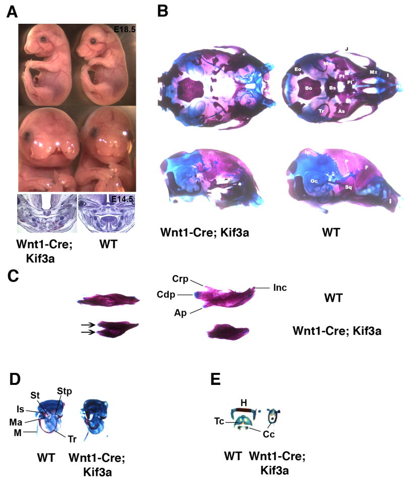
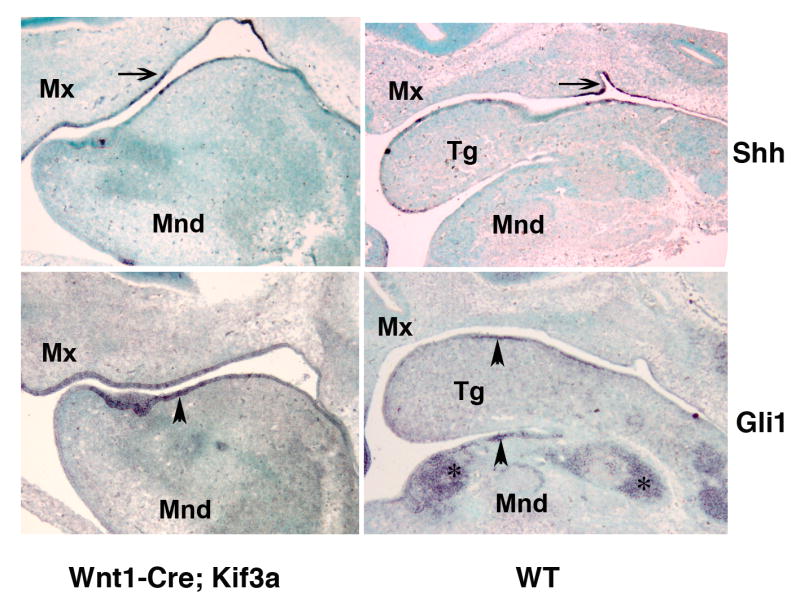
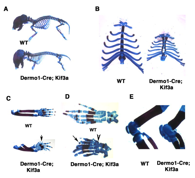
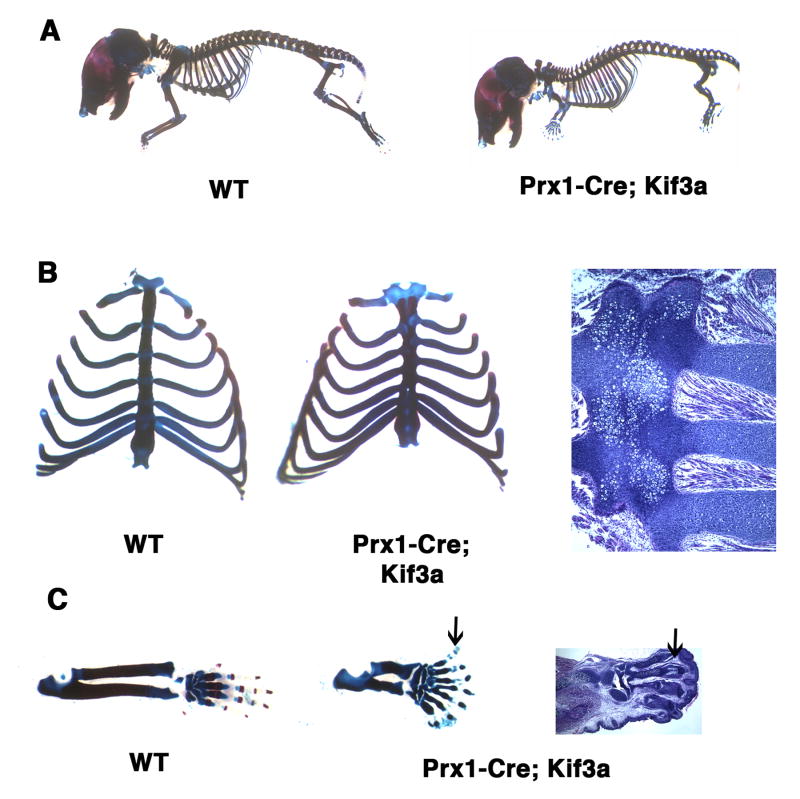
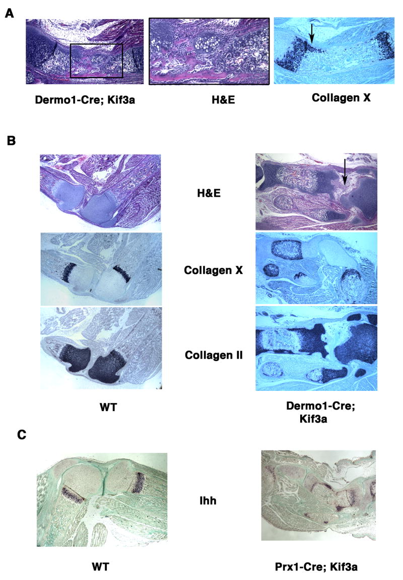
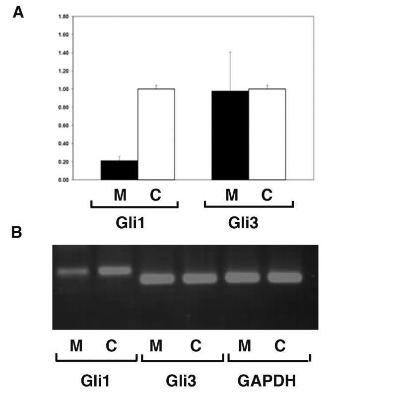
Hedgehog signaling plays an essential role in patterning of the vertebrate skeleton. Here we demonstrate that conditional inactivation of the Kif3a subunit of the kinesin-2 intraflagellar transport motor in mesenchymal skeletal progenitor cells results in severe patterning defects in the craniofacial area, the formation of split sternum and the development of polydactyly. These deformities are reminiscent of those previously described in mice with deregulated hedgehog signaling. We show that in Kif3a-deficient mesenchymal tissues both the repressor function of Gli3 transcription factor and the activation of the Shh transcriptional targets Ptch and Gli1 are compromised. Quantitative analysis of gene expression demonstrates that the Gli1 transcript level is dramatically reduced, whereas Gli3 expression is not significantly affected by kinesin-2 depletion. However, the motor appears to be required for the efficient cleavage of the full-length Gli3 transcription factor into a repressor form.


Figures

Fig. 1
Fig. 1
Craniofacial abnormalities induced by Wnt1-Cre-mediated Kif3a inactivation in neural crest cells. Lateral and frontal view of E18.5 Wnt1-Cre; Kif3a embryos (A, top); H&E staining of frontal sections of E14.5 Wnt1-Cre; Kif3a embryos (A, bottom). Ventral (B, top) and lateral (B, bottom) view of the skull from newborn Wnt1-Cre; Kif3a animal and control littermate stained with Alizarin Red and Alcian Blue. Dorsal (C, left) and medial (C, right) view of mandibles from newborn Wnt1-Cre; Kif3a animal and control littermate. Mutant dentaries are truncated and reveal duplicated lamina (arrows). Otic capsule and middle ear ossicles (D). Tympanic rings in Wnt1-Cre; Kif3a mutants are shortened and display thickening of the lamina. Middle ear ossicles malleus, incus and stapes are absent. Hyoid and laryngeal skeletal elements (E). (Ap) Angular process; (As) alisphenoid; (Bo) basioccipital; (Bs) basisphenoid; (Cc) cricoid cartilage; (Cdp) condylar process; (Crp) coronoid process; (Eo) exoccipital; (F) frontal; (H) hyoid; (I) incisive; (Inc) incisor; (Ip) interparietal; (Is) incus; (J) jugal; (M) Meckel's cartilage; (Ma) malleus; (Mx) maxilla; (Oc) otic capsule; (P) parietal; (Pl) palatine; (Pt) pterygoid; (Sq) squamosal; (St) stapes; (Stp) styloid process; (Tc) thyroid cartilage; (Tr) tympanic ring.
Fig. 2
Fig. 2
Effect of Kif3a ablation on hedgehog signalling in oral epithelium and craniofacial mesenchyme. In situ hybridization of sagittal sections of E12.5 head visualizing Shh (top) and Gli1 (bottom) expression pattern in Wnt1-Cre; Kif3a and control embryos. The gene expression patterns are representative of 5 -7 sagittal sections obtained from E12.5 embryos of each genotype. The expression of Shh (arrow) and its down-stream effector Gli1 (arrowhead) is preserved in Wnt1-Cre; Kif3a mutant and control oral epithelium. In contrast, activation of Gli1 can only be detected in the skeletogenic mesenchyme (asterisks) in the control embryo, whereas it is not evident in the Kif3a-deficient mesenchymal tissue. (Mnd) mandible; (Mx) maxilla; (Tg) tongue.
Fig. 3
Fig. 3
Axial and appendicular skeletal abnormalities induced by genetic ablation of Kif3a in Dermo1-expressing skeletogenic mesoderm. Alizarin Red and Alcian Blue stained skeletal preparations of newborn Dermo1-Cre-mediated Kif3a conditional knockout and control mice. (A-E) Whole mount Dermo1-Cre; Kif3a skeletal preparations demonstrate defects both in axial and appendicular skeleton. The rib cage and long bones of mutant mice are severely reduced in size (A). The Dermo1-Cre; Kif3a mice are characterised by split sternum, thin and shortened ribs (B). The Dermo1-Cre; Kif3a forelimbs display pre-axial polydactyly with duplication of digit I (arrow) and radial hemimelia (C). The Dermo1-Cre; Kif3a hindlimbs develop pre-axial polydactily and digits with missing phalangal elements (arrow). Failure to form joints between embryonic tarsal bones results in multiple bone fusions in newborn limbs (arrowhead) (D). The knee joint display abnormal morphology due to developmental patterning defects of the articular cartilage surfaces of patella, tibia, fibula and femur (E).
Fig. 4
Fig. 4
Sternum and limb defects in newborn mice following lateral plate mesoderm-specific Kif3a inactivation. Whole mount preparations of Prx1-Cre; Kif3a mutants and control newborn mice (A). Mutant fore- and hindlimbs are reduced in length, whereas the rib cage has normal volume (A). The sternal bands in Prx1-Cre; Kif3a mice are fused but slightly shortened, the ribs display normal overall morphology but their attachment sites to sternum are asymmetric (B). The forelimbs of Prx1-Cre; Kif3a mutants develop syndactylous autopods with short dysmorphic digits and ill-defined phalanges (arrows) (C).
Fig. 5
Fig. 5
Effect of kinesin-2 inactivation on the expression of Shh ligand, downstream hedgehog target Ptch and generation of the hedgehog signaling repressor Gli3. At E11.5, expression of Shh transcript is restricted to posterior portion of limb bud in both control and Prx1-Cre; Kif3a embryos (arrows) (A). In response to Shh, expression of Ptch1 gene is up-regulated in posterior domain of both control limb buds and in the mutant hindlimb buds (arrowheads). The transcription level of Ptch1 in the Prx1-Cre; Kif3a forelimb remains low (B). Western blotting of cell lysate from control and Kif3a−/− limb buds demonstrates impaired processing of the full-length Gli3 (Gli3 F) in Kif3a-deficient limbs and decreased amount of the truncated Gli3 repressor (Gli3 Tr) (C).
Fig. 6
Fig. 6
Disorganized growth plate morphology and expression pattern of Ihh in Kif3a-deficient long bones. H&E-stained sections of E18.5 Dermo1-Cre; Kif3a ulna reveals a group of ectopic chondrocytes in the perichondrium (A, left and central panels). In situ hybridization demonstrates abnormal lateral expansion of collagen X-expressing hypertrophic chondrocytes along the affected anterior side of the ulna (A, right panel). H&E staining of paraffin sections of the knee joint area and in situ hybridization with collagen II and collagen-X probes visualize normal epiphyseal distribution of the resting/proliferating and hypertrophic chondrocytes, respectively (B, left panel). In the shortened Kif3a-deficient tibia, an abnormal perichondral distribution of the immature collagen II-positive chondrocytes and hypertrophic chondrocytes is evident (B, right panel). The joint area with articular cartilage defects is marked by arrow. Ihh expression visualized by in situ hybridization is detectable in both control and the Prx1-Cre; Kif3a long bones. Note ectopic expression of Ihh outside the pre-hypertrophic chondrocyte zone of the growth plate in the mutant tibia (C).
Fig. 7
Fig. 7
Kif3 inactivation does not affect Gli3 expression level but prevents up-regulation of Gli1 in long bones. Real-time PCR analysis of total RNA isolated from E18.5 Prx1-Cre; Kif3a and wild type long bones was conducted using Gli1- and Gli3- specific primers. The levels of Gli1 and Gli3 mRNA are calculated relative to GAPDH expression. The mean and SD of the ratios of the Gli1 and Gli3 expression in mutant (M, n=6) vs. control (C, n=6)) are presented (A). All error bars=SD, P<0.001 for Gli1 and P>0.9 for Gli3. The probability values were calculated using two-sample equal variance, two-tailed distribution Student's t-Test. The size and the amount of the Gli1, Gli3 and GAPDH products obtained by RT-PCR were analysed by agarose gel electrophoresis. Gli3 and GAPDH are expressed at comparable level in control and mutant samples, whereas Gli1 expression is markedly reduced in the mutant long bones relative to control (B).

Similar articles

Cited by

References

    1. Bananis E, Nath S, Gordon K, Satir P, Stockert RJ, Murray JW, Wolkoff AW. Microtubule-dependent movement of late endocytic vesicles in vitro: requirements for Dynein and Kinesin. Mol Biol Cell. 2004;15:3688–3697. - PMC - PubMed
    1. Beales PL, Bland E, Tobin JL, Bacchelli C, Tuysuz B, Hill J, Rix S, Pearson CG, Kai M, Hartley J, Johnson C, Irving M, Elcioglu N, Winey M, Tada M, Scambler PJ. IFT80, which encodes a conserved intraflagellar transport protein, is mutated in Jeune asphyxiating thoracic dystrophy. Nat Genet. 2007 In Press. - PubMed
    1. Beech PL, Pagh-Roehl K, Noda Y, Hirokawa N, Burnside B, Rosenbaum JL. Localization of kinesin superfamily proteins to the connecting cilium of fish photoreceptors. J Cell Sci. 1996;109(Pt 4):889–897. - PubMed
    1. Berk M, Desai SY, Heyman HC, Colmenares C. Mice lacking the ski proto-oncogene have defects in neurulation, craniofacial, patterning, and skeletal muscle development. Genes Dev. 1997;11:2029–2039. - PMC - PubMed
    1. Brown CL, Maier KC, Stauber T, Ginkel LM, Wordeman L, Vernos I, Schroer TA. Kinesin-2 is a motor for late endosomes and lysosomes. Traffic. 2005;6:1114–1124. - PubMed

Publication types

MeSH terms

LinkOut - more resources