

A phase II randomized clinical trial of intravitreal bevacizumab for diabetic macular edema
- PMID: 17698196
- PMCID: PMC2245885
- DOI: 10.1016/j.ophtha.2007.05.062
A phase II randomized clinical trial of intravitreal bevacizumab for diabetic macular edema
Abstract
Objective: To provide data on the short-term effect of intravitreal bevacizumab for diabetic macular edema (DME).
Design: Randomized phase II clinical trial.
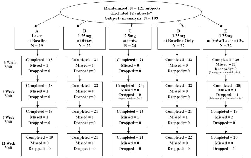
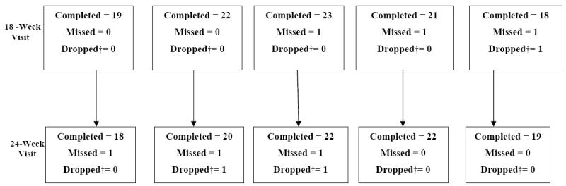
Participants: One hundred twenty-one eyes of 121 subjects (109 eligible for analysis) with DME and Snellen acuity equivalent ranging from 20/32 to 20/320.
Interventions: Random assignment to 1 of 5 groups: (A) focal photocoagulation at baseline (n = 19), (B) intravitreal injection of 1.25 mg of bevacizumab at baseline and 6 weeks (n = 22), (C) intravitreal injection of 2.5 mg of bevacizumab at baseline and 6 weeks (n = 24), (D) intravitreal injection of 1.25 mg of bevacizumab at baseline and sham injection at 6 weeks (n = 22), or (E) intravitreal injection of 1.25 mg of bevacizumab at baseline and 6 weeks with photocoagulation at 3 weeks (n = 22).
Main outcome measures: Central subfield thickness (CST) on optical coherence tomography and best-corrected visual acuity (VA) were measured at baseline and after 3, 6, 9, 12, 18, and 24 weeks.
Results: At baseline, median CST was 411 mum and median Snellen VA equivalent was 20/50. Compared with group A, groups B and C had a greater reduction in CST at 3 weeks and about 1 line better median VA over 12 weeks. There were no meaningful differences between groups B and C in CST reduction or VA improvement. A CST reduction > 11% (reliability limit) was present at 3 weeks in 36 of 84 (43%) bevacizumab-treated eyes and 5 of 18 (28%) eyes treated with laser alone, and at 6 weeks in 31 of 84 (37%) and 9 of 18 (50%) eyes, respectively. Combining focal photocoagulation with bevacizumab resulted in no apparent short-term benefit or adverse outcomes. Endophthalmitis developed in 1 eye. The following events occurred during the first 24 weeks in subjects treated with bevacizumab without attributing cause to the drug: myocardial infarction (n = 2), congestive heart failure (n = 1), elevated blood pressure (n = 3), and worsened renal function (n = 3).
Conclusion: These results demonstrate that intravitreal bevacizumab can reduce DME in some eyes, but the study was not designed to determine whether treatment is beneficial. A phase III trial would be needed for that purpose.
Figures
Similar articles
-
Randomized trial of intravitreal bevacizumab alone or combined with triamcinolone versus macular photocoagulation in diabetic macular edema.Ophthalmology. 2009 Jun;116(6):1142-50. doi: 10.1016/j.ophtha.2009.01.011. Epub 2009 Apr 19. Ophthalmology. 2009. PMID: 19376585 Clinical Trial.
-
Intravitreal bevacizumab (Avastin) therapy for persistent diffuse diabetic macular edema.Retina. 2006 Nov-Dec;26(9):999-1005. doi: 10.1097/01.iae.0000247165.38655.bf. Retina. 2006. PMID: 17151486 Clinical Trial.
-
Association of Baseline Visual Acuity and Retinal Thickness With 1-Year Efficacy of Aflibercept, Bevacizumab, and Ranibizumab for Diabetic Macular Edema.JAMA Ophthalmol. 2016 Feb;134(2):127-34. doi: 10.1001/jamaophthalmol.2015.4599. JAMA Ophthalmol. 2016. PMID: 26605836 Free PMC article. Clinical Trial.
-
Anti-vascular endothelial growth factor for macular oedema secondary to branch retinal vein occlusion.Cochrane Database Syst Rev. 2020 Jul 7;7(7):CD009510. doi: 10.1002/14651858.CD009510.pub3. Cochrane Database Syst Rev. 2020. PMID: 32633861 Free PMC article.
-
Intravitreal steroids for macular edema in diabetes.Cochrane Database Syst Rev. 2020 Nov 17;11(11):CD005656. doi: 10.1002/14651858.CD005656.pub3. Cochrane Database Syst Rev. 2020. PMID: 33206392 Free PMC article.
Cited by
-
Intravitreal Vascular Endothelial Growth Factor Inhibitor Use and Renal Function Decline in Patients with Diabetic Retinopathy.Int J Environ Res Public Health. 2022 Nov 1;19(21):14298. doi: 10.3390/ijerph192114298. Int J Environ Res Public Health. 2022. PMID: 36361175 Free PMC article.
-
Protein kinase cβ phosphorylates occludin regulating tight junction trafficking in vascular endothelial growth factor-induced permeability in vivo.Diabetes. 2012 Jun;61(6):1573-83. doi: 10.2337/db11-1367. Epub 2012 Mar 20. Diabetes. 2012. PMID: 22438576 Free PMC article.
-
Diabetic macular edema: Evidence-based management.Indian J Ophthalmol. 2018 Dec;66(12):1736-1750. doi: 10.4103/ijo.IJO_1240_18. Indian J Ophthalmol. 2018. PMID: 30451174 Free PMC article. Review.
-
Intravitreal diclofenac versus intravitreal bevacizumab in persistent diabetic macular edema: Anatomical and functional outcome.Saudi J Ophthalmol. 2018 Oct-Dec;32(4):303-309. doi: 10.1016/j.sjopt.2018.10.003. Epub 2018 Oct 13. Saudi J Ophthalmol. 2018. PMID: 30581301 Free PMC article.
-
Long-term therapeutic efficacy of the subthreshold micropulse diode laser photocoagulation for diabetic macular edema.Jpn J Ophthalmol. 2011 Jul;55(4):365-369. doi: 10.1007/s10384-011-0033-3. Epub 2011 Jun 7. Jpn J Ophthalmol. 2011. PMID: 21647567
References
-
- Early Treatment Diabetic Retinopathy Study Research Group. Photocoagulation for diabetic macular edema: Early Treatment Diabetic Retinopathy Study report number 1. Arch Ophthalmol. 1985;103:1796–806. - PubMed
-
- Early Treatment Diabetic Retinopathy Study Research Group. Early photocoagulation for diabetic retinopathy: ETDRS report number 9. Ophthalmology. 1991;98(suppl):766–85. - PubMed
-
- Diabetes Control and Complication Trial Research Group. The effect of intensive treatment of diabetes on the development and progression of long-term complications in insulin-dependent diabetes mellitus. N Engl J Med. 1993;329:977–86. - PubMed
-
- UK Prospective Diabetes Study (UKPDS) Group. Risks of progression of retinopathy and vision loss related to tight blood pressure control in type 2 diabetes mellitus: UKPDS 69. Arch Ophthalmol. 2004;122:1631–40. - PubMed
-
- Martidis A, Duker JS, Greenberg PB, et al. Intravitreal triamcinolone for refractory diabetic macular edema. Ophthalmology. 2002;109:920–7. - PubMed
Publication types
MeSH terms
Substances
Grants and funding
LinkOut - more resources
Full Text Sources
Other Literature Sources
Medical
