

doi: 10.1016/j.jbiotec.2007.07.498.
Epub 2007 Jul 12.
A non-isotopic in vitro assay for histone acetylation
Affiliations
- PMID: 17698235
- PMCID: PMC2099255
- DOI: 10.1016/j.jbiotec.2007.07.498
Item in Clipboard
A non-isotopic in vitro assay for histone acetylation
J Biotechnol.
.
Abstract
We describe a simple, robust, and relatively inexpensive non-radioactive in vitro assay for measuring histone acetyl-transferase activity. The assay takes advantage of easy to purify recombinant E. coli-derived fusion proteins containing the NH(2)-terminal tails of histones H3 and H4 linked to epitope-tagged maltose-binding protein (MBP), and immunoblotting with antibodies specific to acetylated H3 and H4. Here we show the specificity and dynamic range of this assay for the histone acetyl-transferases, p300 and PCAF. This assay may be adapted readily for other substrates by simply generating new fusion proteins and for other acetyl-transferases by modifying reaction conditions.
Figures

A. Diagram illustrating the linear structure of fusion proteins containing the NH2-terminal part of Tetrahymena histone H3 (amino acids 1 – 22) or H4 (residues 1 – 19) joined to maltose binding protein (MBP), followed by a flag epitope. Lysine resides in each histone segment are indicated with bold type. Positions of 5’ and 3’ multiple cloning sites (mcs) also are indicated. B. Purification of E. coli-derived H3- and H4-MBP. Pictured is an image of a stained SDS-PAGE gel of soluble E. coli protein lysates before and after induction of the fusion gene with IPTG (U - un-induced, I - induced), and following purification of the chimeric proteins using amylose resin (P). For details see “Materials and Methods”.

A. Dose-dependent acetylation of purified H4-MBP (upper panels) after incubation for 45 min with p300 immunoprecipitated from 50 µg of nuclear protein extracts from C3H10T1/2 cells infected with Ad-p300 (lower panels). Equivalent amounts of MBP or H4-MBP proteins were used (middle panels). Results from 2 independent experiments are displayed to the right in graphical form. B. Kinetics of H4-MBP acetylation by p300. Time course studies show increases in H4 acetylation (upper panel) after incubation of 0.5 µg of purified H4-MBP fusion protein (middle panel) for 0 – 240 min with Flag-tagged p300 immunoprecipitated from 50 µg of C3H10T1/2 cell nuclear protein extracts (lower panels). Results of 3 independent experiments (mean ± SEM) are graphed to the right. C. Saturable acetylation of H4-MBP by p300. Flag-tagged p300 was isolated by immunoprecipitation from different amounts of nuclear protein extracts (NE) obtained from C3H10T1/2 cells infected with Ad-p300 (upper panel), and was incubated with 0.5 µg of H4-MBP (middle panel) for 45 min, followed by analysis of acetylated H4 by immunoblotting (lower panel). Results from 2 independent experiments are graphed to the right.
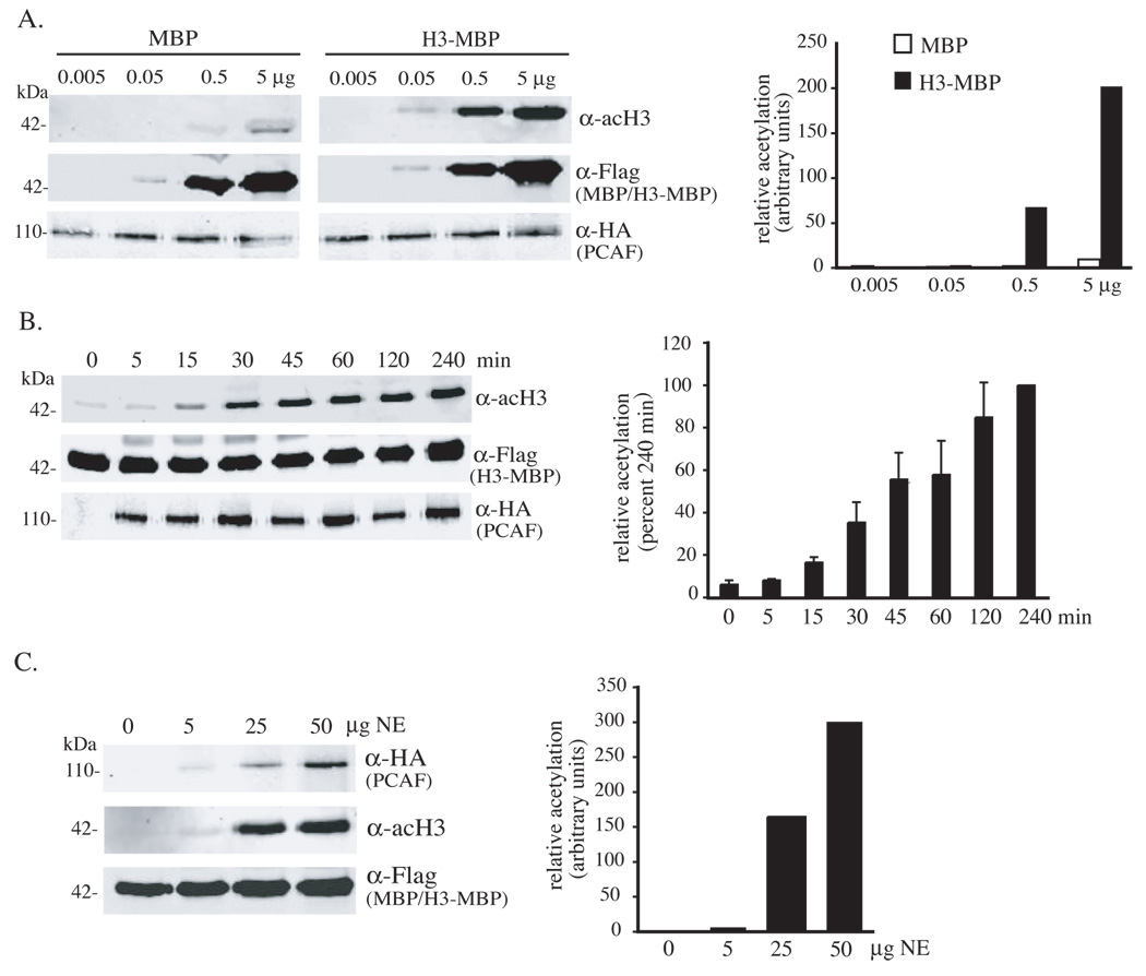
A. Dose-dependent acetylation of purified H3-MBP (upper panels) after incubation for 45 min with HA-tagged PCAF immunoprecipitated from 50 µg of nuclear protein extracts from C3H10T1/2 cells infected with Ad-PCAF (lower panels). Equivalent amounts of MBP and H3-MBP proteins were used (middle panels). Results from 2 independent experiments are displayed to the right. B. Kinetics of H3-MBP acetylation by PCAF. Time course studies demonstrate increases in H4 acetylation (upper panel) after incubation of 0.5 µg of purified H3-MBP fusion protein (middle panel) with HA-tagged PCAF immunoprecipitated from 50 µg of C3H10T1/2 cell nuclear protein extracts (lower panels). Results of 3 independent experiments (mean ± SEM) are graphed to the right. C. Saturable acetylation of H3-MBP by PCAF. HA-tagged PCAF was isolated by immunoprecipitation from different amounts of NE from C3H10T1/2 cells infected with Ad-PCAF (upper panel), and was incubated with 0.5 µg of H3-MBP (middle panel) for 45 min. Acetylated H3 was assessed by immunoblotting (lower panel). Results from 2 independent experiments are displayed to the right.

A. Comparison of acetylation of MBP, H3-MBP, and H4-MBP (upper panels) by p300 and PCAF, and by an E. coli-derived GST-PCAF fusion protein. Equivalent amounts of each fusion protein (0.5 µg) were used in each assay (lower panels). B. Recombinant p300 with mutations in the acetyltransferase domain (ATmut) cannot acetylate H4-MBP (upper panel). Identical amounts of H4-MBP (0.5 µg) were used in each experiment (middle panels), and concentrations of wild type and mutant p300 were equivalent (lower panels). C. Recombinant PCAF with mutations in the acetyltransferase domain (ATmut) cannot acetylate H3-MBP (upper panel). Identical amounts of H3-MBP (0.5 µg) were used in each experiment (middle panels), and concentrations of wild type (wt) and mutant PCAF were comparable (lower panels).
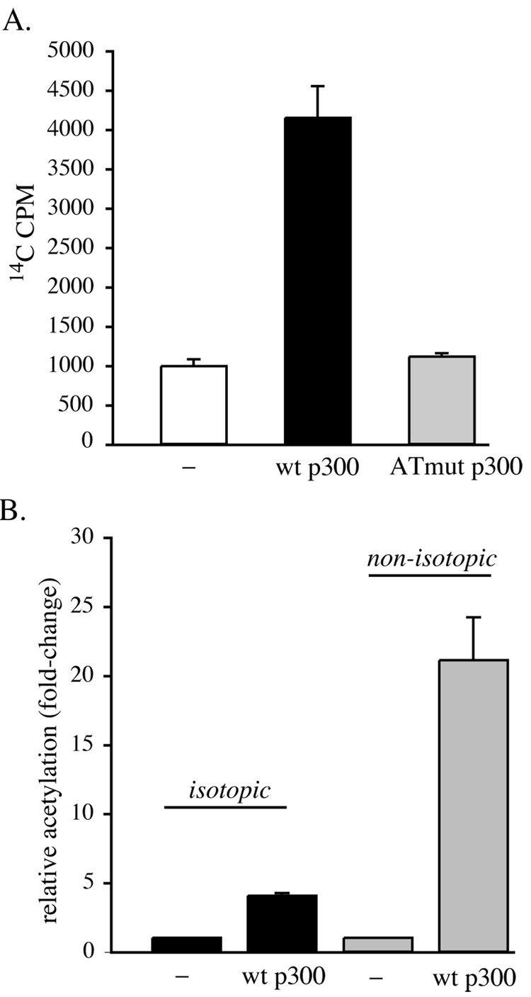
A. Core histones (10 µg) were incubated with immunoprecipitated wild-type (wt) p300, the acetyltransferase domain mutant (ATmut), or no p300 (−) for 45 min in the presence of 14C-acetyl CoA, as described in “Materials and Methods”. Results of 3 independent experiments (mean ± SEM) are graphed. B. Comparison of isotopic acetylation of 10 µg of core histones by p300 with non-isotopic labeling of 0.5 µg of H4-MBP. Equivalent amounts of immunoprecipitated p300 were used in each group of experiments. Results of 3 independent assays (mean ± SEM) are depicted.
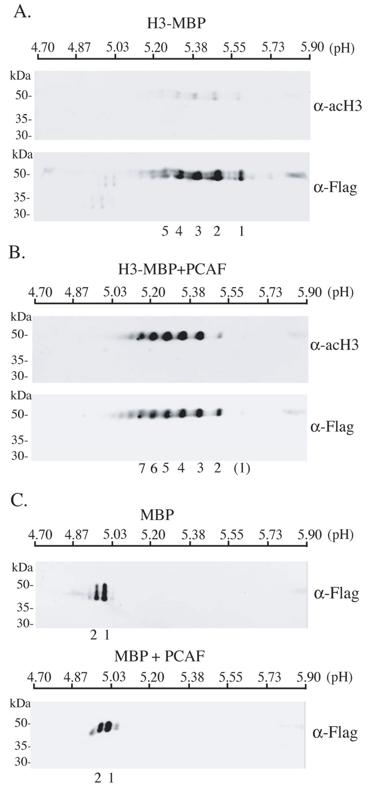
A. Detection of H3-MBP (0.5 µg) by immunoblotting using anti-acH3 (upper panel) or anti-Flag antibodies (lower panel) after isoelectric focusing using linear pH 4.7 to 5.9 gradient gels and SDS-PAGE. B. Detection of H3-MBP (0.5 µg) by immunoblotting using anti-acH3 (upper panel) or anti-Flag antibodies (lower panel) after incubation with GST-PCAF, followed by isoelectric focusing and SDS-PAGE, as described in A. C. Detection of MBP by immunoblotting using anti-Flag antibodies before (upper panel) or after (lower panel) incubation with GST-PCAF, and followed by isoelectric focusing and SDS-PAGE, as described in A. For A – C, individual spots are numbered. The numbers correspond to one another in A and B, and in both parts of C.
References
-
- Barrera LO, Ren B. The transcriptional regulatory code of eukaryotic cells--insights from genome-wide analysis of chromatin organization and transcription factor binding. Curr. Opin. Cell Biol. 2006;18:291–298. - PubMed
-
- Berndsen CE, Denu JM. Assays for mechanistic investigations of protein/histone acetyltransferases. Methods. 2005;36:321–331. - PubMed
-
- Herrera JE, Bergel M, Yang XJ, Nakatani Y, Bustin M. The histone acetyltransferase activity of human GCN5 and PCAF is stabilized by coenzymes. J. Biol. Chem. 1997;272:27253–27258. - PubMed
-
- Kalkhoven E. CBP and p300: HATs for different occasions. Biochem. Pharmacol. 2004;68:1145–1155. - PubMed
Publication types
MeSH terms
Substances
Grants and funding
LinkOut - more resources
Full Text Sources
Other Literature Sources
Miscellaneous
