

Diffusion tensor imaging in children with periventricular leukomalacia: variability of injuries to white matter tracts
- PMID: 17698519
- PMCID: PMC7977654
- DOI: 10.3174/ajnr.A0534
Diffusion tensor imaging in children with periventricular leukomalacia: variability of injuries to white matter tracts
Abstract
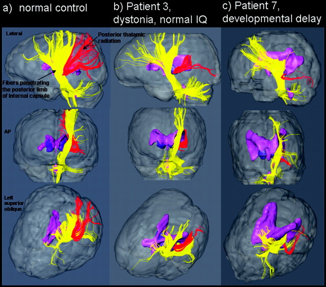
Background and purpose: Conventional MR imaging shows evidence of brain injury and/or maldevelopment in 70%-90% of children with cerebral palsy (CP), though its capability to identify specific white matter tract injury is limited. The great variability of white matter lesions in CP already demonstrated by postmortem studies is thought to be one of the reasons why response to treatment is so variable. Our hypothesis is that diffusion tensor imaging (DTI) is a suitable technique to provide in vivo characterization of specific white matter tract lesions in children with CP associated with periventricular leukomalacia (PVL).
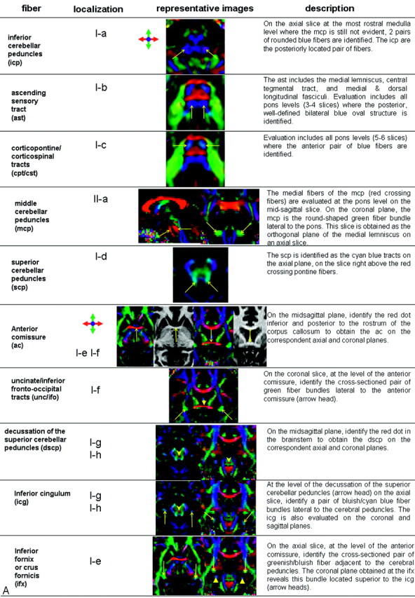
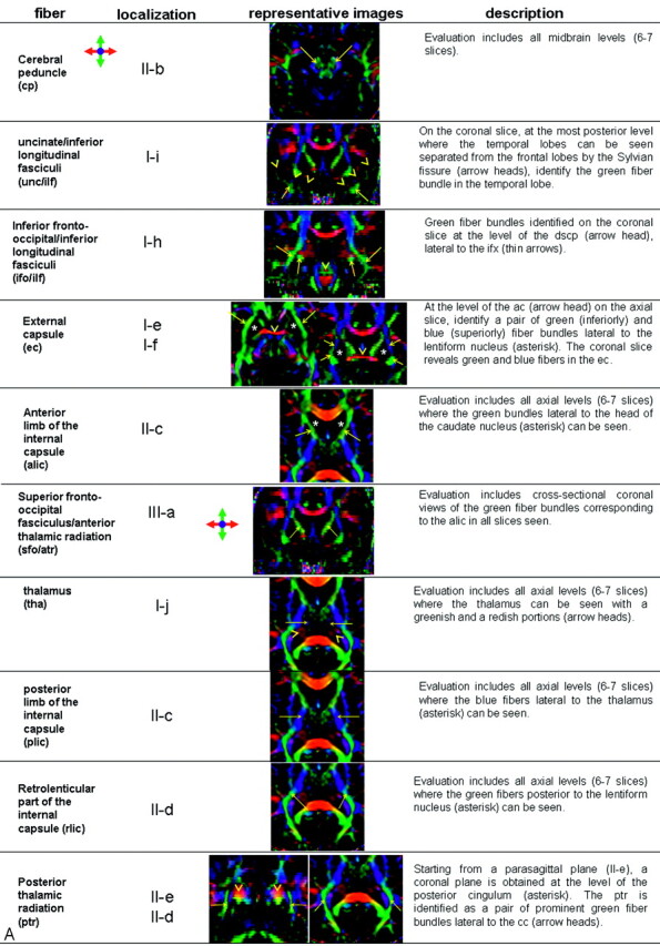
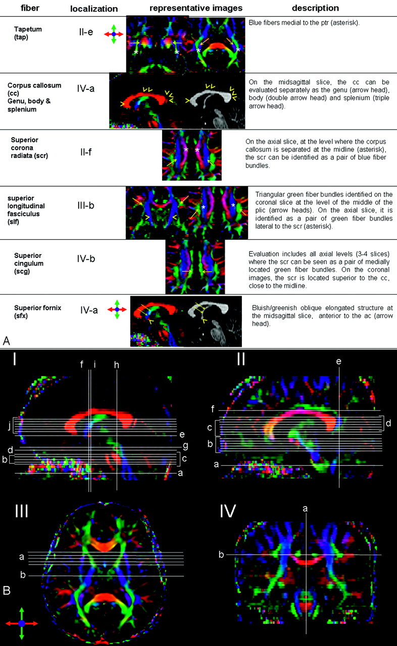
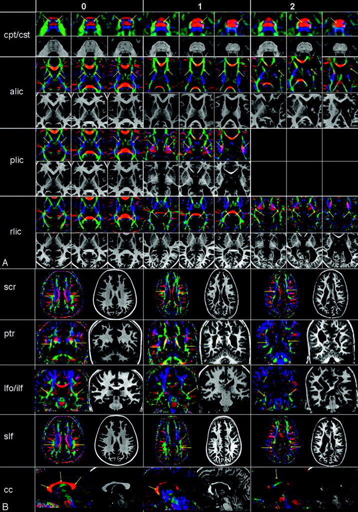
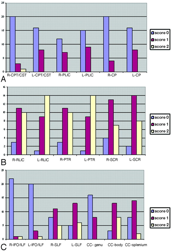
Materials and methods: In this study, 24 children with CP associated with PVL and 35 healthy controls were evaluated with DTI. Criteria for identification of 26 white matter tracts on the basis of 2D DTI color-coded maps were established, and a qualitative scoring system, based on visual inspection of the tracts in comparison with age-matched controls, was used to grade the severity of abnormalities. An ordinal grading system (0=normal, 1=abnormal, 2=severely abnormal or absent) was used to score each white matter tract.
Results: There was marked variability in white matter injury pattern in patients with PVL, with the most frequent injury to the retrolenticular part of the internal capsule, posterior thalamic radiation, superior corona radiata, and commissural fibers.
Conclusion: DTI is a suitable technique for in vivo assessment of specific white matter lesions in patients with PVL and, thus, a potentially valuable diagnostic tool. The tract-specific evaluation revealed a family of tracts that are highly susceptible in PVL, important information that can potentially be used to tailor treatment options in the future.
Figures
References
-
- Osler SW. The Cerebral Palsies of Children. London, UK: Mac Keith Press;1987
-
- Keogh JM, Badawi N. The origins of cerebral palsy. Curr Opin Neurol 2006;19:129–34 - PubMed
-
- Volpe JJ. Cerebral white matter injury of the premature infant: more common that you think. Pediatrics 2003;112:176–80 - PubMed
-
- Folkerth RD. Neuropathologic substrate of cerebral palsy. J Child Neurol 2005;20:940–49 - PubMed
Publication types
MeSH terms
Grants and funding
LinkOut - more resources
Full Text Sources
Medical
Miscellaneous