

High-resolution contrast-enhanced, susceptibility-weighted MR imaging at 3T in patients with brain tumors: correlation with positron-emission tomography and histopathologic findings
- PMID: 17698528
- PMCID: PMC7977663
- DOI: 10.3174/ajnr.A0540
High-resolution contrast-enhanced, susceptibility-weighted MR imaging at 3T in patients with brain tumors: correlation with positron-emission tomography and histopathologic findings
Abstract
Background and purpose: The purpose of this work was to demonstrate susceptibility effects (SusE) in various types of brain tumors with 3T high-resolution (HR)-contrast-enhanced (CE)-susceptibility-weighted (SW)-MR imaging and to correlate SusE with positron-emission tomography (PET) and histopathology.
Materials and methods: Eighteen patients with brain tumors, scheduled for biopsy or tumor extirpation, underwent high-field (3T) MR imaging. In all of the patients, an axial T1-spin-echo (SE) sequence and an HR-SW imaging sequence before and after IV application of a standard dose of contrast agent (MultiHance) was obtained. Seven patients preoperatively underwent PET. The frequency and formation of intralesional SusE in all of the images were evaluated and correlated with tumor grade as determined by PET and histopathology. Direct correlation of SusE and histopathologic specimens was performed in 6 patients. Contrast enhancement of the lesions was assessed in both sequences.
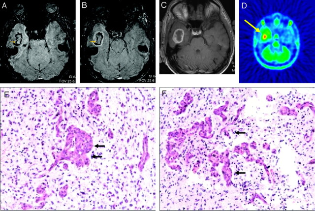
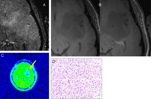
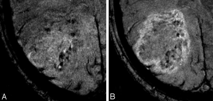
Results: High-grade lesions demonstrated either high or medium frequency of SusE in 90% of the patients. Low-grade lesions demonstrated low frequency of SusE or no SusE. Correlation between intralesional frequency of SusE and histopathologic, as well as PET, tumor grading was statistically significant. Contrast enhancement was equally visible in both SW and SE sequences. Side-to-side comparison of tumor areas with high frequency of SusE and histopathology revealed that intralesional SusE reflected conglomerates of increased tumor microvascularity.
Conclusions: 3T HR-CE-SW-MR imaging shows both intratumoral SusE not visible with standard MR imaging and contrast enhancement visible with standard MR imaging. Because frequency of intratumoral SusE correlates with tumor grade as determined by PET and histopathology, this novel technique is a promising tool for noninvasive differentiation of low-grade from high-grade brain tumors and for determination of an optimal area of biopsy for accurate tumor grading.
Figures
Similar articles
-
High-field, high-resolution, susceptibility-weighted magnetic resonance imaging: improved image quality by addition of contrast agent and higher field strength in patients with brain tumors.Neuroradiology. 2008 Jan;50(1):9-16. doi: 10.1007/s00234-007-0298-x. Epub 2007 Sep 18. Neuroradiology. 2008. PMID: 17876570
-
Improved preoperative evaluation of cerebral cavernomas by high-field, high-resolution susceptibility-weighted magnetic resonance imaging at 3 Tesla: comparison with standard (1.5 T) magnetic resonance imaging and correlation with histopathological findings--preliminary results.Invest Radiol. 2007 Jun;42(6):346-51. doi: 10.1097/01.rli.0000262744.85397.fc. Invest Radiol. 2007. PMID: 17507804
-
Glial tumor grading and outcome prediction using dynamic spin-echo MR susceptibility mapping compared with conventional contrast-enhanced MR: confounding effect of elevated rCBV of oligodendrogliomas [corrected].AJNR Am J Neuroradiol. 2004 Feb;25(2):214-21. AJNR Am J Neuroradiol. 2004. PMID: 14970020 Free PMC article.
-
The advantage of high relaxivity contrast agents in brain perfusion.Eur Radiol. 2006 Nov;16 Suppl 7:M16-26. doi: 10.1007/s10406-006-0192-3. Eur Radiol. 2006. PMID: 18655263 Review.
-
MultiHance in brain tumor imaging.Eur Radiol. 2004 Jun;14 Suppl 7:O5-9; discussion O20-1. doi: 10.1007/s10406-004-0057-6. Eur Radiol. 2004. PMID: 15503370 Review. No abstract available.
Cited by
-
Combination of high-resolution susceptibility-weighted imaging and the apparent diffusion coefficient: added value to brain tumour imaging and clinical feasibility of non-contrast MRI at 3 T.Br J Radiol. 2010 Jun;83(990):466-75. doi: 10.1259/bjr/34304111. Epub 2009 Aug 18. Br J Radiol. 2010. PMID: 19690076 Free PMC article.
-
Emerging Techniques in Brain Tumor Imaging: What Radiologists Need to Know.Korean J Radiol. 2016 Sep-Oct;17(5):598-619. doi: 10.3348/kjr.2016.17.5.598. Epub 2016 Aug 23. Korean J Radiol. 2016. PMID: 27587949 Free PMC article. Review.
-
MRI grading versus histology: predicting survival of World Health Organization grade II-IV astrocytomas.AJNR Am J Neuroradiol. 2015 Jan;36(1):77-83. doi: 10.3174/ajnr.A4077. Epub 2014 Aug 7. AJNR Am J Neuroradiol. 2015. PMID: 25104288 Free PMC article.
-
Susceptibility Imaging in Glial Tumor Grading; Using 3 Tesla Magnetic Resonance (MR) System and 32 Channel Head Coil.Pol J Radiol. 2017 Apr 1;82:179-187. doi: 10.12659/PJR.900374. eCollection 2017. Pol J Radiol. 2017. PMID: 28439322 Free PMC article.
-
MR imaging detection of cerebral microbleeds: effect of susceptibility-weighted imaging, section thickness, and field strength.AJNR Am J Neuroradiol. 2009 Feb;30(2):338-43. doi: 10.3174/ajnr.A1355. Epub 2008 Nov 11. AJNR Am J Neuroradiol. 2009. PMID: 19001544 Free PMC article. Clinical Trial.
References
-
- Barth M, Nobauer-Huhmann IM, Reichenbach JR, et al. High-resolution three-dimensional contrast-enhanced blood oxygenation level-dependent magnetic resonance venography of brain tumors at 3 Tesla: first clinical experience and comparison with 1.5 Tesla. Invest Radiol 2003;38:409–14 - PubMed
-
- Rauscher A, Sedlacik J, Fitzek C, et al. High resolution susceptibility weighted MR-imaging of brain tumors during the application of a gaseous agent. Rofo 2005;177:1065–69 - PubMed
-
- Reichenbach JR, Barth M, Haacke EM, et al. High-resolution MR venography at 3.0 Tesla. J Comput Assist Tomogr 2000;24:949–57 - PubMed
-
- Reichenbach JR, Essig M, Haacke EM, et al. High-resolution venography of the brain using magnetic resonance imaging. Magma 1998;6:62–69 - PubMed
MeSH terms
Substances
LinkOut - more resources
Full Text Sources
Medical