

Increasing contrast agent concentration improves enhancement in first-pass CT perfusion
- PMID: 17698531
- PMCID: PMC7977633
- DOI: 10.3174/ajnr.A0574
Increasing contrast agent concentration improves enhancement in first-pass CT perfusion
Abstract
Background and purpose: Our aim was to evaluate whether increasing iodine concentration, at a constant total iodine dose, resulted in better brain tissue opacification in patients with acute stroke symptoms during their evaluation by first-pass CT perfusion (CTP).
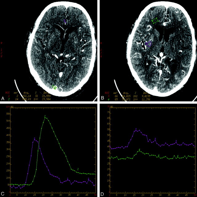
Materials and methods: One hundred two patients presenting to the emergency department within 3 hours of onset of acute stroke symptoms underwent CTP scanning. Three different concentrations of iodinated nonionic contrast material were used (300, 350, or 400 mg/mL). Total iodine dose (15 g) and injection rate (7 mL/s) were kept constant. There were 25, 53, and 19 patients in the different concentration groups, respectively; 5 patients were excluded due to uncorrectable motion artifacts. CTP scanning was performed at the level of the putamen, and data were analyzed by determining peak opacification for normal gray and white matter, arterial input, and venous output. Mean and SD values were calculated, and 3 concentration groups, stratified by region-of-interest location, were compared by using a single-tailed unpaired t test.
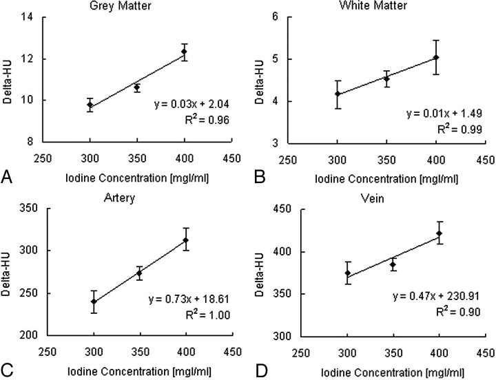
Results: Monotonic increasing peak opacification was observed in all region-of-interest locations. Statistically significant differences were observed between the 300 and 350 mg/mL, 300 and 400 mg/mL, as well as the 350 and 400 mg/mL groups (P<.01) in white matter, gray matter, and the arterial input. Statistical significance was seen in the venous output group between the 300 and 400 mg/mL (P<.005) and 350 and 400 mg/mL (P<.007) groups, but not between the 300 and 350 mg/mL (P=.2) groups.
Conclusion: Increasing contrast concentration improves peak opacification of tissue, suggesting that CTP evaluation of patients with acute stroke is better performed with the highest available concentration contrast agent.
Figures
Similar articles
-
Intravascular enhancement with identical iodine delivery rate using different iodine contrast media in a circulation phantom.Invest Radiol. 2013 Nov;48(11):813-8. doi: 10.1097/RLI.0b013e31829979e8. Invest Radiol. 2013. PMID: 23857135
-
Which Iodine concentration in chest CT?--a prospective study in 300 patients.Eur Radiol. 2008 Dec;18(12):2826-32. doi: 10.1007/s00330-008-1080-0. Epub 2008 Jul 24. Eur Radiol. 2008. PMID: 18651154
-
Optimization of contrast material delivery for dual-energy computed tomography pulmonary angiography in patients with suspected pulmonary embolism.Invest Radiol. 2012 Jan;47(1):78-84. doi: 10.1097/RLI.0b013e31821a2142. Invest Radiol. 2012. PMID: 21577132 Clinical Trial.
-
Contrast timing in computed tomography: effect of different contrast media concentrations on bolus geometry.Eur J Radiol. 2012 Apr;81(4):e629-32. doi: 10.1016/j.ejrad.2011.12.038. Epub 2012 Jan 31. Eur J Radiol. 2012. PMID: 22297184
-
Multiphase contrast-enhanced CT of the liver with a multislice CT scanner: effects of iodine concentration and delivery rate.Radiat Med. 2005 Feb;23(1):61-9. Radiat Med. 2005. PMID: 15786754 Clinical Trial.
Cited by
-
Contrast enhancement efficacy of iodinated contrast media: Effect of molecular structure on contrast enhancement.Eur J Radiol Open. 2018 Oct 6;5:183-188. doi: 10.1016/j.ejro.2018.09.005. eCollection 2018. Eur J Radiol Open. 2018. PMID: 30310828 Free PMC article.
-
Role of Permeability Surface Area Product in Grading of Brain Gliomas using CT Perfusion.Asian J Neurosurg. 2023 Nov 7;18(4):751-760. doi: 10.1055/s-0043-1774820. eCollection 2023 Dec. Asian J Neurosurg. 2023. PMID: 38161609 Free PMC article.
-
Effects of different tube potentials and iodine concentrations on image enhancement, contrast-to-noise ratio and noise in micro-CT images: a phantom study.Quant Imaging Med Surg. 2013 Oct;3(5):256-61. doi: 10.3978/j.issn.2223-4292.2013.10.04. Quant Imaging Med Surg. 2013. PMID: 24273743 Free PMC article.
-
Predictors of hyperperfusion syndrome after stent implantation in symptomatic intracranial atherosclerotic stenosis.Quant Imaging Med Surg. 2023 Feb 1;13(2):1048-1057. doi: 10.21037/qims-22-682. Epub 2022 Dec 19. Quant Imaging Med Surg. 2023. PMID: 36819235 Free PMC article.
-
Biased visualization of hypoperfused tissue by computed tomography due to short imaging duration: improved classification by image down-sampling and vascular models.Eur Radiol. 2015 Jul;25(7):2080-8. doi: 10.1007/s00330-015-3602-x. Epub 2015 Apr 17. Eur Radiol. 2015. PMID: 25894005
References
-
- Latchaw RE, Yonas H, Hunter GJ, et al. Guidelines and recommendations for perfusion imaging in cerebral ischemia: a scientific statement for health care professionals by the writing group on perfusion imaging from the Council on Cardiovascular Radiology of the American Heart Association. Stroke 2003;34:1084–104 - PubMed
-
- Esteban JM, Cervera V. Perfusion CT and angio CT in assessment of acute stroke. Neuroradiology 2004;46:705–15 - PubMed
-
- Eastwood JD, Lev MH, Provenzale JM. Perfusion CT with iodinated contrast material. AJR Am J Roentgenol 2003;180:3–12 - PubMed
Publication types
MeSH terms
Substances
LinkOut - more resources
Full Text Sources
Medical