Comparative Study
. 2007 Aug;28(7):1328-31.
doi: 10.3174/ajnr.A0544.

Wernicke encephalopathy: MR findings at clinical presentation in twenty-six alcoholic and nonalcoholic patients

Affiliations
Comparative Study

Wernicke encephalopathy: MR findings at clinical presentation in twenty-six alcoholic and nonalcoholic patients

G Zuccoli et al. AJNR Am J Neuroradiol. 2007 Aug.

Abstract

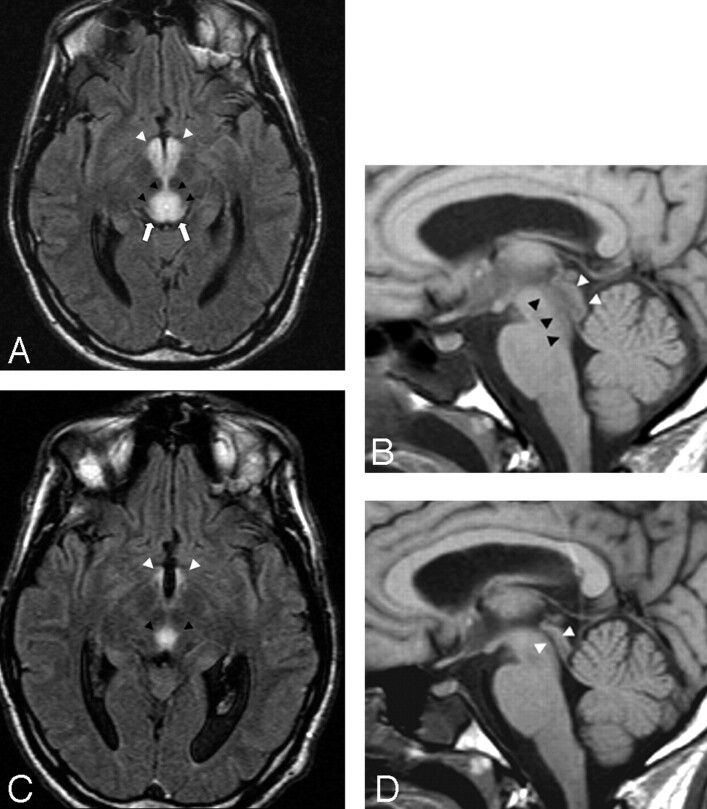
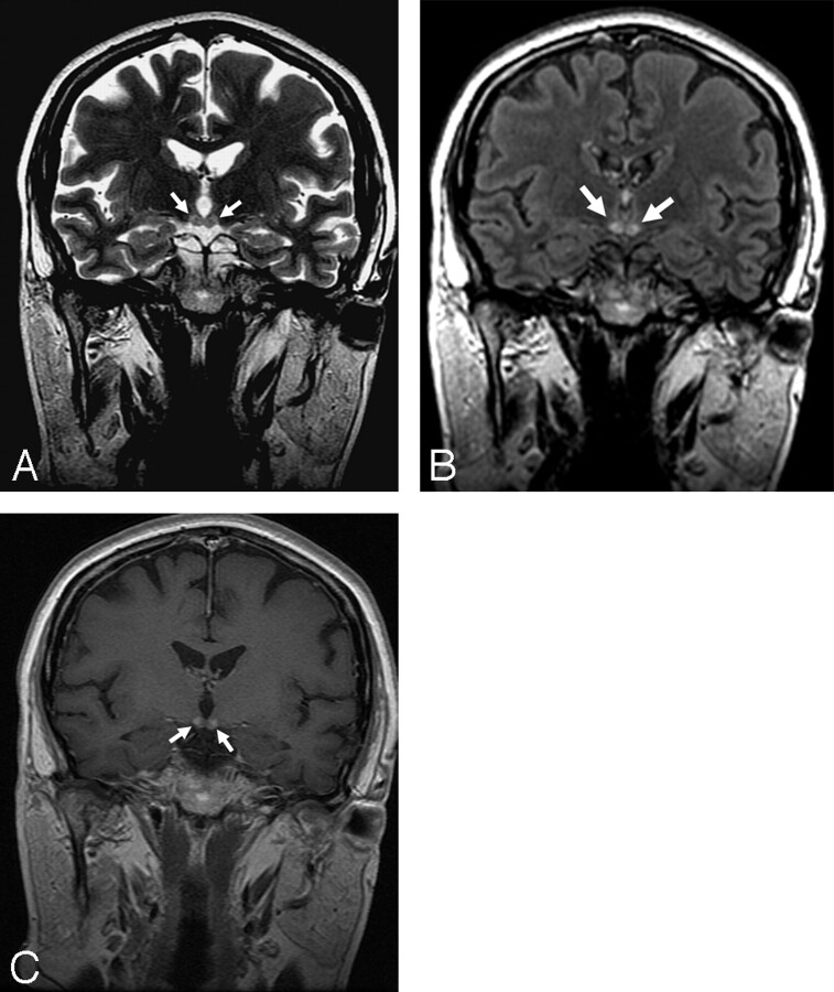
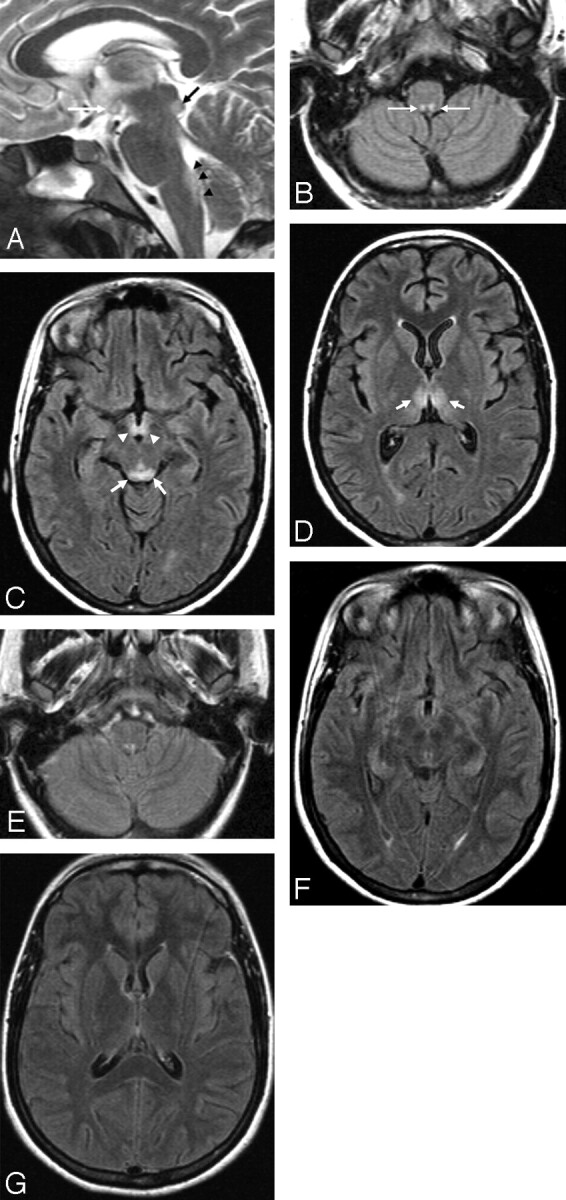
Background and purpose: Wernicke encephalopathy is a severe neurologic disorder that results from a dietary vitamin B1 deficiency. It is characterized by changes in consciousness, ocular abnormalities, and ataxia. This study was undertaken to analyze and compare findings on MR imaging and neurologic symptoms at clinical presentations of patients with Wernicke encephalopathy with and without a history of alcohol abuse.

Materials and methods: A multicenter study group retrospectively reviewed MR brain imaging findings, clinical histories, and presentations of 26 patients (14 female, 12 male) diagnosed between 1999 and 2006 with Wernicke encephalopathy. The age range was 6-81 years (mean age, 46 .6+/-19 years).

Results: Fifty percent of the patients had a history of alcohol abuse, and 50% had no history of alcohol abuse. Eighty percent showed changes in consciousness, 77% had ocular symptoms, and 54% had ataxia. Only 38% of the patients showed the classic triad of the disease at clinical presentation. At MR examination, 85% of the patients showed symmetric lesions in the medial thalami and the periventricular region of the third ventricle, 65% in the periaqueductal area, 58% in the mamillary bodies, 38% in the tectal plate, and 8% in the dorsal medulla. Contrast enhancement of the mamillary bodies was statistically positively correlated with the alcohol abuse group.

Conclusions: Our study confirms the usefulness of MR in reaching a prompt diagnosis of Wernicke encephalopathy to avoid irreversible damage to brain tissue. Contrast enhancement in the mamillary bodies is a typical finding of the disease in the alcoholic population.


Figures

Fig 1.
Fig 1.
A 33-year-old man presented with sudden and progressive changes in consciousness after prolonged voluntary food starvation. A, Axial FLAIR (11000/140/2 [TR/TE/NEX]) image shows marked hyperintensity of the tectal region (white arrows), periaqueductal area (black arrowheads), and mamillary bodies (white arrowheads). The lesions are compatible with reversible cytotoxic edema. B, On sagittal T1-weighted (530/13/2 [TR/TE/NEX]) image, low signal intensity areas with respect to the pons of both periaqueductal areas (black arrowheads) and tectal plate (white arrowheads) are seen. Note that the local swelling determines mild stenosis of the cerebral aqueduct. C, Ten days after thiamine replacement, a partial regression of the lesions involving the aqueductal region (black arrowheads) and mamillary bodies (white arrowheads) is easily seen on axial FLAIR image. D, This finding is confirmed by a sagittal T1-weighted image showing reopening of the cerebral aqueduct (white arrowheads).
Fig 2.
Fig 2.
A 53-year-old woman with an history of chronic alcohol abuse presented with the classic neurologic triad of Wernicke encephalopathy. Coronal T2-weighted (2500/90/2 [TR/TE/NEX]) (A) and coronal FLAIR images (9000/105/2 [TR/TE/NEX]) (B) show high signal intensity circumscribed to the mamillary bodies (white arrows). After administration of contrast media, central enhancement (white arrows) of both mamillary bodies is seen on coronal T1-weighted image (532/15/2 [TR/TE/NEX]) (C).
Fig 3.
Fig 3.
A 21-year-old woman presented with hyperemesis gravidarum, changes in consciousness, and ocular abnormalities. A, Sagittal T2-weighted image (4447/100/4 [TR/TE/NEX]) shows high signal intensity of the lower tectal plate (black arrow), dorsal upper medulla (black arrowheads), and mamillary body (white arrow). B, Symmetric involvement of the prepositus hypoglossal nuclei (white arrows) is demonstrated on an axial FLAIR (11000/140/2 [TR/TE/NEX]) image. C, Alterations in the tectal plate (white arrows) and mamillary bodies (white arrowheads) are seen. D, Axial FLAIR image at the level of the basal ganglia shows alterations of the medial thalamic nuclei (white arrows). E-G, Twelve days after the start of thiamine replacement therapy, regression of neurologic symptoms and FLAIR abnormalities is seen.
Fig 4.
Fig 4.
A 45-year-old woman with a history of alcohol abuse had changes in consciousness and ocular abnormalities. A, No signal intensity alterations are seen on axial T2-weighted (2500/90/2 [TR/TE/NEX]) image at the level of the mamillary bodies and periaqueductal area. No alterations are seen on T2-weighted image at the level of the basal ganglia (not shown). B, After administration of contrast media, contrast enhancement of the mamillary bodies is demonstrated (white arrows) on axial T1-weighted (532/15/2 [TR/TE/NEX]) image.

Similar articles

Cited by

References

    1. Harper CG, Giles M, Finlay-Jones R. Clinical signs in the Wernicke-Korsakoff complex: a retrospective analysis of 131 cases diagnosed at necropsy. J Neurol Neurosurg Psychiatry 1986;49:341–45 - PMC - PubMed
    1. Ogershok PR, Rahman A, Nestor S, et al. Wernicke encephalopathy in nonalcoholic patients. Am J Med Sci 2002;323:107–11 - PubMed
    1. Gallucci M, Bozzao A, Splendiani A, et al. Wernicke encephalopathy: MR findings in five patients. AJNR Am J Neuroradiol 1990;11:887–92 - PMC - PubMed
    1. Victor M. The Wernicke-Korsakoff syndrome. In: Vinken PJ, Bruyn GW, eds. Handbook of Clinical Neurology. Vol 28 . Amsterdam: North Holland;1976. :243–70
    1. Harper C, Butterworth R. Nutritional and metabolic disorders. In: Graham DI, Lantos PL, eds. Greenfield's Neuropathology,vol 1 , 6th ed. London: Hodder Arnold;1997. :601–52