

Incorporating functional MR imaging into diffusion tensor tractography in the preoperative assessment of the corticospinal tract in patients with brain tumors
- PMID: 17698540
- PMCID: PMC7977658
- DOI: 10.3174/ajnr.A0538
Incorporating functional MR imaging into diffusion tensor tractography in the preoperative assessment of the corticospinal tract in patients with brain tumors
Abstract
Background and purpose: Our goal was to improve the preoperative assessment of the corticospinal tract (CST) in patients with brain tumors. We investigated whether the integration of functional MR imaging (fMRI) data and diffusion tensor (DT) tractography can be used to evaluate the spatial relationship between the hand and foot fibers of the CST and tumor borders.
Materials and methods: We imaged 10 subjects: 1 healthy volunteer and 9 patients. Imaging consisted of a 3D T1-weighted sequence, a gradient-echo echo-planar imaging (EPI) sequence for fMRI, and a diffusion-weighted EPI sequence for DT tractography. DT tractography was initiated from a seed region of interest in the white matter area subjacent to the maximal fMRI activity in the precentral cortex. The target region of interest was placed in the cerebral peduncle.
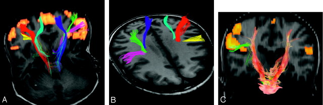
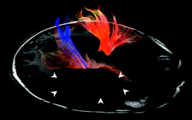
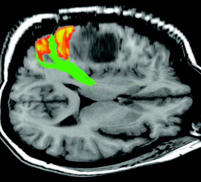
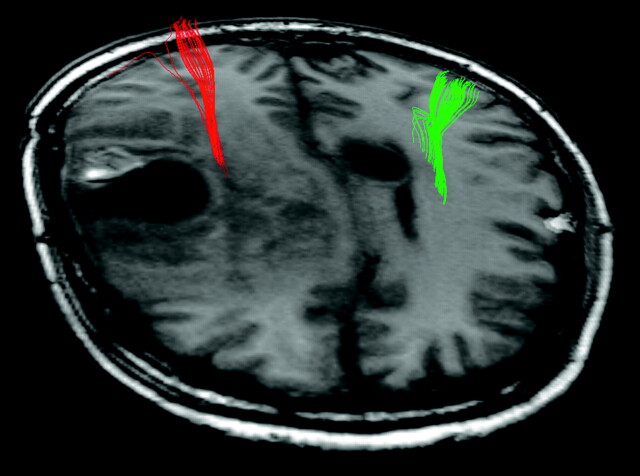
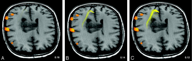
Results: In the healthy volunteer, we successfully tracked hand, foot, and lip fibers bilaterally by using fMRI-based DT tractography. In all patients, we could track the hand fibers of the CST bilaterally. In 4 patients who also performed foot tapping, we could clearly distinguish hand and foot fibers. We were able to depict the displacement of hand and foot fibers by tumor and the course of fibers through areas of altered signal intensity.
Conclusion: Incorporating fMRI into DT tractography in the preoperative assessment of patients with brain tumors may provide additional information on the course of important white matter tracts and their relationship to the tumor. Only this approach allows a distinction between the CST components, while visualization of the CST is improved when fiber tracking is hampered by tumor (infiltration) or perifocal edema.
Figures
References
-
- Sunaert S, Yousry TA. Clinical applications of functional magnetic resonance imaging. Neuroimaging Clin N Am 2001;11:221–36, viii - PubMed
-
- Haughton VM, Turski PA, Meyerand B, et al. The clinical applications of functional MR imaging. Neuroimaging Clin N Am 1999;9:285–93 - PubMed
-
- Yousry TA, Schmid UD, Jassoy AG, et al. Topography of the cortical motor hand area: prospective study with functional MR imaging and direct motor mapping at surgery. Radiology 1995;195:23–29 - PubMed
-
- Pujol J, Conesa G, Deus J, et al. Presurgical identification of the primary sensorimotor cortex by functional magnetic resonance imaging. J Neurosurg 1996;84:7–13 - PubMed
Publication types
MeSH terms
LinkOut - more resources
Full Text Sources
Other Literature Sources
Medical