. 2007 Oct;75(10):5043-51.
doi: 10.1128/IAI.00508-07. Epub 2007 Aug 13.

Prolonged colonization of mice by Vibrio cholerae El Tor O1 depends on accessory toxins

Affiliations

Prolonged colonization of mice by Vibrio cholerae El Tor O1 depends on accessory toxins

Verena Olivier et al. Infect Immun. 2007 Oct.

Abstract

Cholera epidemics caused by Vibrio cholerae El Tor O1 strains are typified by a large number of asymptomatic carriers who excrete vibrios but do not develop diarrhea. This carriage state was important for the spread of the seventh cholera pandemic as the bacterium was mobilized geographically, allowing the global dispersion of this less virulent strain. Virulence factors associated with the development of the carriage state have not been previously identified. We have developed an animal model of cholera in adult C57BL/6 mice wherein V. cholerae colonizes the mucus layer and forms microcolonies in the crypts of the distal small bowel. Colonization occurred 1 to 3 h after oral inoculation and peaked at 10 to 12 h, when bacterial loads exceeded the inoculum by 10- to 200-fold, indicating bacterial growth within the small intestine. After a clearance phase, the number of bacteria within the small intestine, but not those in the cecum or colon, stabilized and persisted for at least 72 h. The ability of V. cholerae to prevent clearance and establish this prolonged colonization was associated with the accessory toxins hemolysin, the multifunctional autoprocessing RTX toxin, and hemagglutinin/protease and did not require cholera toxin or toxin-coregulated pili. The defect in colonization attributed to the loss of the accessory toxins may be extracellularly complemented by inoculation of the defective strain with an isogenic colonization-proficient V. cholerae strain. This work thus demonstrates that secreted accessory toxins modify the host environment to enable prolonged colonization of the small intestine in the absence of overt disease symptoms and thereby contribute to disease dissemination via asymptomatic carriers.


Figures

FIG. 1.
FIG. 1.
Colonization dynamics of V. cholerae. (A to C) C57BL/6 mice were inoculated with 1.5 × 10⁶ CFU of either wild-type (wt) P27459 (A), CT mutant strain P4 (B), or multitoxin-deficient mutant strain KFV101 (C). (D) Comparison of median values for P27459 (filled diamonds), P4 (filled triangles), and KFV101 (unfilled squares). After 1, 3, 6, 12, 24, 48, and 72 h, small intestines (sm. int.; trisected into proximal [prox.], middle [mid.], and distal [dist.] portions), ceca, and colons were collected, homogenized, and plated for CFU counting. Individual and median values are displayed as log colonization indices (col. ind.; log numbers of CFU recovered/log numbers of CFU inoculated). The experiment was performed twice with six to eight mice per group. Numbers below data points refer to mice from whom the numbers of CFU recovered were below the detection limit (d.l.; dotted lines and corresponding arrows at the bottoms of the panels). Solid lines and corresponding arrows indicate the inoculum (i). At some time points, similar colonization indices were obtained for different mice, and consequently, the diamonds, triangles, or squares are superimposed.
FIG. 2.
FIG. 2.
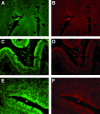
V. cholerae was found in the crypts of the small intestines. Mice were inoculated with 108 CFU of wild-type P27459, and after 6 h, the intestines were collected and 1-cm-thick sections were fixed in Carnoy's fixative. Bacteria were labeled by FISH and identified as V. cholerae by using a probe specific for Vibrio 16S RNA (A, C, and E) and/or eubacteria (B, D, and F). Bacteria were found in the crypts of the small intestinal villi (A and B), in the lumina (C and D), and on top of Peyer's patches (E and F).
FIG. 3.
FIG. 3.
TCP were not required for colonization of adult mice. Mice were inoculated with 5 × 10⁶ to 10 × 106 CFU of wild-type V. cholerae P27459 (filled diamonds) or 5 × 10⁶ CFU of the ΔtcpA mutant (unfilled diamonds). After 12, 20, and 48 h, the mice were sacrificed and the small intestines were collected, homogenized, and plated for CFU counting. Individual and median values are displayed as log colonization indices (col. ind.; log numbers of CFU recovered/log numbers of CFU inoculated). The arrow on the left and the corresponding dotted line indicate the detection limit (d.l.); the arrow on the right and the corresponding solid line indicate the inoculum (i). At some time points, similar colonization indices were obtained for different mice, and consequently, the diamonds are superimposed.
FIG. 4.
FIG. 4.
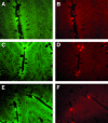
TCP were not required for microcolony formation. Mice were inoculated with 108 CFU of either wild-type P27459 (A and B) or the ΔtcpA mutant (C to F). Sections of the ileocecal junctions were fixed in Carnoy's fixative 6 hpi. FISH labeling of sections was performed using a Vibrio-specific probe (A, C, and E) and a probe labeling eubacteria (B, D, and F).
FIG. 5.
FIG. 5.
KFV101 colonized the small intestines of adult mice when inoculated along with P4. Mice were inoculated with various mixtures of P4 and KFV101, and 48 hpi, homogenized small intestines were plated for CFU counting. Individual and median values are displayed as log colonization indices (col. ind.; log numbers of CFU recovered/log numbers of CFU inoculated) for P4 (triangles), KFV101 alone (unfilled square), and P4 and KFV101 in combination (comb.; filled squares). Numbers below data points refer to mice from whom the numbers of CFU recovered were below the detection limit (d.l.; lower arrow and corresponding dotted line). The upper arrow and corresponding solid line indicate the inoculum (i). At some time points, similar colonization indices were obtained for different mice, and consequently, the triangles or squares are superimposed.

Similar articles

Cited by

References

    1. Anderson, A. M., J. B. Varkey, C. A. Petti, R. A. Liddle, R. Frothingham, and C. W. Woods. 2004. Non-O1 Vibrio cholerae septicemia: case report, discussion of literature, and relevance to bioterrorism. Diagn. Microbiol. Infect. Dis. 49:295-297. - PubMed
    1. Angelichio, M. J., J. Spector, M. K. Waldor, and A. Camilli. 1999. Vibrio cholerae intestinal population dynamics in the suckling mouse model of infection. Infect. Immun. 67:3733-3739. - PMC - PubMed
    1. Bart, K. J., Z. Huq, M. Khan, and W. H. Mosley. 1970. Seroepidemiologic studies during a simultaneous epidemic of infection with El Tor Ogawa and classical Inaba Vibrio cholerae. J. Infect. Dis. 121(Suppl.):S17-S24. - PubMed
    1. Barua, D. 1992. History of cholera, p.1-36. In D. Barua and W. B. Greenough III (ed.), Cholera. Plenum Medical Book Company, New York, NY.
    1. Blake, P. A., R. E. Weaver, and D. G. Hollis. 1980. Diseases of humans (other than cholera) caused by vibrios. Annu. Rev. Microbiol. 34:341-367. - PubMed

Publication types

MeSH terms

LinkOut - more resources