. 2007 Oct;27(20):7063-72.
doi: 10.1128/MCB.00769-07. Epub 2007 Aug 13.

Species selectivity of mixed-lineage leukemia/trithorax and HCF proteolytic maturation pathways

Affiliations

Species selectivity of mixed-lineage leukemia/trithorax and HCF proteolytic maturation pathways

Francesca Capotosti et al. Mol Cell Biol. 2007 Oct.

Abstract

Site-specific proteolytic processing plays important roles in the regulation of cellular activities. The histone modification activity of the human trithorax group mixed-lineage leukemia (MLL) protein and the cell cycle regulatory activity of the cell proliferation factor herpes simplex virus host cell factor 1 (HCF-1) are stimulated by cleavage of precursors that generates stable heterodimeric complexes. MLL is processed by a protease called taspase 1, whereas the precise mechanisms of HCF-1 maturation are unclear, although they are known to depend on a series of sequence repeats called HCF-1(PRO) repeats. We demonstrate here that the Drosophila homologs of MLL and HCF-1, called Trithorax and dHCF, are both cleaved by Drosophila taspase 1. Although highly related, the human and Drosophila taspase 1 proteins display cognate species specificity. Thus, human taspase 1 preferentially cleaves MLL and Drosophila taspase 1 preferentially cleaves Trithorax, consistent with coevolution of taspase 1 and MLL/Trithorax proteins. HCF proteins display even greater species-specific divergence in processing: whereas dHCF is cleaved by the Drosophila taspase 1, human and mouse HCF-1 maturation is taspase 1 independent. Instead, human and Xenopus HCF-1PRO repeats are cleaved in vitro by a human proteolytic activity with novel properties. Thus, from insects to humans, HCF proteins have conserved proteolytic maturation but evolved different mechanisms.


Figures

FIG. 1.
FIG. 1.
Human and Drosophila MLL/Trx and HCF proteins. (A) Schematic structures of human MLL and Drosophila Trx proteins. Architectural elements are identified above the schematic: PHD, plant homeodomain; FYRN, MLLN carboxy-terminal association element; FYRC, MLLC amino-terminal association element; SET, histone H3 lysine 4 methyltransferase domain. The positions of the two taspase 1 cleavage sites (CS1 and CS2) are indicated as red and yellow arrowheads, respectively. Conserved regions in Trx are shown as for MLL. The line labeled E3 indicates the region deleted in the mutant TrxE3 protein. (B) Schematic structures of human HCF-1 and dHCF proteins. Architectural elements are identified above the schematic. HCF-1KEL, Kelch repeat domain; Basic and Acidic, regions enriched in basic and acidic residues, respectively; HCF-1PRO, HCF-1 proteolytic processing repeats; Fn3, fibronectin type 3 repeats; NLS, nuclear localization signal. Conserved regions in dHCF are shown in the same colors; basic and acidic regions that display similar charge bias but not sequence identity are shown in related colors. The position of the dHCF CS1-like taspase 1 recognition site is indicated by the arrowhead and dashed line.
FIG. 2.
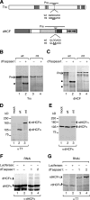
FIG. 2.
Trx and dHCF are cleaved by Drosophila taspase 1. (A) Schematic of Trx and dHCF cleavage precursors. The lines labeled Pre indicate the region contained within each precursor. Wild-type (wt) and mutant (mt) versions of the putative taspase 1 cleavage sites are shown below. Note that the two diagrams are not drawn to the same scale. (B) Drosophila taspase 1 (dTaspase1) proteolytic activity on the Trx precursor. 35S-labeled wild-type (lanes 1 and 2) or CS2-like mutant (lanes 3 and 4) Trx precursors were incubated for 2 h at 37°C with (lanes 2 and 4) or without (lanes 1 and 3) purified recombinant Drosophila taspase 1. Products were resolved by SDS-PAGE and revealed by autoradiography. •, N-terminal cleavage product. A smaller C-terminal fragment is not visible owing to the reduced specific activity of this product. (C) Drosophila taspase 1 proteolytic activity on the dHCF precursor. 35S-labeled wild-type (lanes 1 and 2) or CS1-like mutant (lanes 3 and 4) dHCF precursors were incubated with (lanes 2 and 4) or without (lanes 1 and 3) purified recombinant Drosophila taspase 1. Products were resolved by SDS-PAGE and revealed by autoradiography. •, location of larger N-terminal and smaller C-terminal cleavage products. (D) dHCF cleavage at the Drosophila taspase 1 cleavage site in vivo. SL2 cells were mock transfected (lane 1) or transfected with wild-type (lane 2) or CS1-like mutant (lane 3) T7-tagged full-length dHCF expression vector. Proteolysis by endogenous protease was assessed by anti-T7 tag (αT7) immunoblotting. rdHCFFL, full-length rdHCF; rdHCFN, rdHCF N-terminal subunit. Molecular mass markers are listed on the left. (E) Anti-dHCFN antibody (αdHCFN) reveals processing of endogenous dHCF. Endogenous dHCF proteolysis of the samples shown in panel D was revealed by immunoblot analysis with affinity-purified anti-dHCFN antibody. (F) RNAi depletion of Drosophila taspase 1 impairs endogenous dHCF processing. SL2 cells were mock treated (lane 1) or treated with luciferase (lane 2) or two independent taspase 1 dsRNAs (RNAi 1 in lane 3 and RNAi 2 in lane 4) for 48 h before cleavage analysis by anti-dHCFN immunoblotting. (G) RNAi depletion of Drosophila taspase 1 impairs dHCF processing. SL2 cells were treated with the indicated dsRNAs as in panel F for 48 h before transfection of the T7-tagged full-length dHCF expression vector. dHCF cleavage was analyzed 48 h after the transfection by anti-T7 immunoblotting.
FIG. 3.
FIG. 3.
Coevolution of human and Drosophila taspase 1 with their specific substrates. (A) Human taspase 1 versus Drosophila taspase 1 proteolytic activity on dHCF cleavage. Wild-type dHCF precursor was incubated without extract (lanes 1 and 4) or with HeLa (lane 2) and SL2 (lane 3) cytosolic extracts or recombinant human taspase 1 (lane 5) and Drosophila taspase 1 (lane 6). (B) Schematic of MLL and Trx cleavage precursors. The line labeled Pre indicates the region contained within each precursor. The Trx(MLL CS2) precursor contains the Trx cleavage site (QMDGVDD) changed to the MLL CS2 site sequence (QLDGVDD). (C) Human and Drosophila cell extract activities on MLL and Trx cleavage. MLL (lanes 1 to 3) and Trx (lanes 4 to 6) precursors were incubated without extract (lanes 1 and 4) or with HeLa (lanes 2 and 5) or SL2 (lanes 3 and 6) cytosolic extracts. (D) Human taspase 1 and Drosophila taspase 1 activities on MLL and Trx cleavage. MLL (lanes 1 to 3) and Trx (lanes 4 to 6) precursors were incubated without taspase 1 (lanes 1 and 4) or with recombinant human taspase 1 (lanes 2 and 5) or Drosophila taspase 1 (lanes 3 and 6). (E) Human taspase 1 and Drosophila taspase 1 activities on humanized Trx(MLLCS2) precursor. The Trx(MLLCS2) precursor was incubated without taspase 1 (lane 1) or with recombinant human taspase 1 (lane 2) or Drosophila taspase 1 (lane 3). For all cleavage products, black dots indicate the N-terminal cleavage products; smaller, lower-specific-activity C-terminal fragments are not visible.
FIG. 4.
FIG. 4.
HCF-1 is not cleaved by taspase 1. (A) Schematic of HCF-1 cleavage precursor. The line labeled Pre indicates the region contained within the precursor. Wild-type (wt) and mutated (mt) versions of the HCF-1PRO repeat 1 are shown below. (B) HCF-1PRO repeats are not evident substrates for human taspase 1 (hTaspase1) cleavage. 35S-labeled wild-type (lanes 1, 2, 5, and 6) or mutated (lanes 3, 4, 7, and 8) HCF-1 and MLL precursors were incubated with (lanes 2, 4, 6, and 8) or without (lanes 1, 3, 5, and 7) purified recombinant human taspase 1. •, larger N-terminal and shorter C-terminal cleavage products. (C) taspase 1−/− cells contain normally processed HCF-1. Wild-type (lane 2) and taspase 1−/− (lane 3) MEF lysates were resolved by 7% SDS-PAGE. Cleavage of endogenous HCF-1 was detected by anti-HCF-1C (αHCF-1C) immunoblot analysis. HeLa nuclear extract (lane 1) was used as an HCF-1 cleavage pattern control. —, HCF-1 precursor; •, HCF-1PRO repeat cleavage products.
FIG. 5.
FIG. 5.
HCF-1PRO repeats are faithfully processed by human cell extracts. (A) Schematic of HCF-1 cleavage precursors. The line labeled Pre indicates the region contained within each precursor. These HCF-1 precursors contain the first three HCF-1PRO repeats in a wild-type version (HCF-1rep123) or with mutated repeat 1 (HCF-1repX23), mutated repeat 2 (HCF-1rep1X3), or mutated repeats 1 and 2 (HCF-1repXX3). Products corresponding to full-length precursor (Pre) and N-terminal cleavage products for HCF-1PRO repeats 3, 2, and 1 are shown below. (B) HeLa cell extract cleavage of HCF-1PRO repeats. 35S-labeled HCF-1 precursors from panel A were incubated without (lanes 1, 3, 5, and 7) or with (lanes 2, 4, 6, and 8) nuclear HeLa cell extract. Numbers on the right of each panel indicate the N-terminal cleavage product derived from the indicated HCF-1PRO repeat; x indicates the missing cleavage product corresponding to the mutated HCF-1PRO repeat; C-terminal fragments are not visible owing to their reduced specific activity. (C) Time course of the HeLa cell extract proteolytic activity. 35S-labeled HCF-1rep123 precursor was incubated with nuclear HeLa cell extracts for the indicated periods of time. (D) Quantification of time course data. The relative accumulation of HCF-1PRO repeat 1 cleavage product from panel C was quantified to represent HCF-1PRO repeat cleavage activity over time. (E) Characterization of the HeLa cell proteolytic activity. 35S-labeled HCF-1rep123 precursor was incubated without extract (lane 1) or with nuclear HeLa cell extract (lanes 2 to 5) that was heat treated (lane 2), untreated (lane 3), or treated with Pefabloc (lane 4) or Complete protease inhibitor (PI) mixture (lane 5).
FIG. 6.
FIG. 6.
Evolution of HCF protein maturation. (A) HCF protein maturation in insects. Top, schematic representation of dHCF. The taspase 1 cleavage site is indicated by the arrowhead. Bottom, charge profiles of the fly Drosophila melanogaster and the honeybee Apis mellifera HCF proteins. Peaks above zero indicate basic regions, and peaks below zero indicate acidic regions; basic and acidic regions are shown in blue and red, respectively. For each protein, sequences of taspase 1 cleavage sites are indicated above arrowheads. (B) HCF protein maturation in vertebrates. Top, schematic representation of human HCF-1. Segments corresponding to the basic and acidic regions and to the HCF-1PRO repeats are indicated above the schematic. Bottom, charge profiles of the human Homo sapiens, frog Xenopus tropicalis, and fish Fugus rubripes HCF proteins as in panel A. For each protein, the region corresponding to the HCF-1PRO repeats is overlined. Sequences corresponding to partially conserved taspase 1 cleavage sites are indicated above arrowheads; in red is indicated the residue that does not match the taspase 1 site consensus. (C) Sequence conservation of the HCF-1PRO repeats in vertebrates. An alignment of fish (top), frog (center), and human (bottom) HCF-1PRO repeats is shown. The human HCF-1PRO repeats are numbered as in reference . Residues matching a consensus based on the most frequent residue at each position are shaded. Positions in the consensus sequences for which a conserved residue cannot be defined are indicated by dashes. Underlined positions indicate sequence conservation among HCF-1PRO repeats of all three species. •, residues important for human HCF-1PRO repeat cleavage in vivo (32). (D) In vitro cleavage of xHCF-1. Top, schematic representation of xHCF-1. The line labeled Pre indicates the region contained in the cleavage precursor. The dashed line indicates the imperfect taspase 1 cleavage site. Bottom, xHCF-1 precursor (xHCF-1rep89) was incubated without additions (lane 4) or with either Drosophila taspase 1 (dTaspase1) (lane 5), human taspase 1 (hTaspase1) (lane 6), or HeLa cell extract (lane 7). dHCF (lanes 1 to 3) and human HCF-1 (hHCF-1rep123; lanes 8 and 9) precursors are shown as positive controls for taspase 1 (dHCF) and HCF-1PRO repeat (hHCF-1rep123) cleavage. •, HCF protein cleavage products.

References

    1. Ayton, P. M., and M. L. Cleary. 2001. Molecular mechanisms of leukemogenesis mediated by MLL fusion proteins. Oncogene 20:5695-5707. - PubMed
    1. Breen, T. R., and P. J. Harte. 1991. Molecular characterization of the trithorax gene, a positive regulator of homeotic gene expression in Drosophila. Mech. Dev. 35:113-127. - PubMed
    1. Brown, M. S., and J. L. Goldstein. 1997. The SREBP pathway: regulation of cholesterol metabolism by proteolysis of a membrane-bound transcription factor. Cell 89:331-340. - PubMed
    1. Dignam, J. D., R. M. Lebovitz, and R. G. Roeder. 1983. Accurate transcription initiation by RNA polymerase II in a soluble extract from isolated mammalian nuclei. Nucleic Acids Res. 11:1475-1489. - PMC - PubMed
    1. Djabali, M., L. Selleri, P. Parry, M. Bower, B. D. Young, and G. A. Evans. 1992. A trithorax-like gene is interrupted by chromosome 11q23 translocations in acute leukaemias. Nat. Genet. 2:113-118. - PubMed

Publication types

MeSH terms

LinkOut - more resources