

Cellular effects of reversed amidines on Trypanosoma cruzi
- PMID: 17698624
- PMCID: PMC2151434
- DOI: 10.1128/AAC.00047-07
Cellular effects of reversed amidines on Trypanosoma cruzi
Abstract
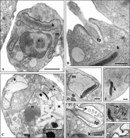
Aromatic diamidines represent a class of DNA minor groove-binding ligands that exhibit high levels of antiparasitic activity. Since the chemotherapy for Chagas' disease is still an unsolved problem and previous reports on diamidines and related analogues show that they have high levels of activity against Trypanosoma cruzi infection both in vitro and in vivo, our present aim was to evaluate the cellular effects in vitro of three reversed amidines (DB889, DB702, and DB786) and one diguanidine (DB711) against both amastigotes and bloodstream trypomastigotes of T. cruzi, the etiological agent of Chagas' disease. Our data show that the reversed amidines have higher levels of activity than the diguanidine, with the order of trypanocidal activities being as follows: DB889 > DB702 > DB786 > DB711. Transmission electron microscopy analysis showed that the reversed amidines induced many alterations in the nuclear morphology, swelling of the endoplasmic reticulum and Golgi structures, and consistent damage in the mitochondria and kinetoplasts of the parasites. Interestingly, in trypomastigotes treated with the reversed amidine DB889, multiple axoneme structures (flagellar microtubules) were noted. Flow cytometry analysis confirmed that the treated parasites presented an important loss of the mitochondrial membrane potential, as revealed by a decrease in rhodamine 123 fluorescence. Our results show that the reversed amidines have promising activities against the relevant mammalian forms of T. cruzi and display high trypanocidal effects at very low doses. This is especially the case for DB889, which merits further in vivo evaluation.
Figures


References
-
- Araujo-Jorge, T. C., E. P. Sampaio, W. De Souza, and M. N. Meirelles. 1989. Trypanosoma cruzi: the effect of variations in experimental conditions on the levels of macrophage infection in vitro. Parasitol. Res. 75:257-263. - PubMed
-
- Baselin, M., M. A. Badet-Denisot, and M. Robert-Gero. 1998. Modification of kinetoplast DNA minicircle composition in pentamidine-resistant Leishmania. Acta Trop. 70:43-61. - PubMed
-
- Braga, M. V., and W. De Souza. 2006. Effects of protein kinase and phosphatidylinositol-3 kinase inhibitors on growth and ultrastructure of Trypanosoma cruzi. FEMS Microbiol. Lett. 256:209-216. - PubMed
-
- Coura, R. J., and S. L. De Castro. 2002. A critical review on Chagas' disease chemotherapy. Mem. Inst. Oswaldo Cruz 97:3-24. - PubMed
-
- Croft, S. L., and R. P. Brazil. 1982. Effect of pentamidine isethionate on the ultrastructure and morphology of Leishmania mexicana amazonensis in vitro. Ann. Trop. Med. Parasitol. 76:37-43. - PubMed
Publication types
MeSH terms
Substances
LinkOut - more resources
Full Text Sources
