Patterns of atrophy differ among specific subtypes of mild cognitive impairment
- PMID: 17698703
- PMCID: PMC2735186
- DOI: 10.1001/archneur.64.8.1130
Patterns of atrophy differ among specific subtypes of mild cognitive impairment
Abstract
Background: In most patients, mild cognitive impairment (MCI) represents the clinically evident prodromal phase of dementia. This is most well established in amnestic MCI, which is most commonly a precursor to Alzheimer disease (AD). It follows, however, that subjects with MCI who have impairment in nonmemory domains may progress to non-AD degenerative dementias.
Objective: To investigate patterns of cerebral atrophy associated with specific subtypes of MCI.
Design: Case-control study.
Setting: Community-based sample at a tertiary referral center.
Patients: One hundred forty-five subjects with MCI and 145 age- and sex-matched cognitively normal control subjects. Mild cognitive impairment was classified as amnestic, single cognitive domain; amnestic, multiple domain; nonamnestic, single domain; and nonamnestic, multiple domain. Subjects with nonamnestic single-domain MCI were classified into language, attention/executive, and visuospatial subgroups on the basis of specific cognitive impairment.
Main outcome measure: Patterns of gray matter loss in the MCI groups compared with control subjects, assessed using voxel-based morphometry.
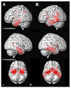
Results: Subjects in the amnestic single- and multiple-domain groups showed loss in the medial and inferior temporal lobes compared with control subjects, and those in the multiple-domain group also had involvement of the posterior temporal lobe, parietal association cortex, and posterior cingulate. Subjects in the nonamnestic single-domain group with language impairment showed loss in the left anterior inferior temporal lobe. The group with attention/executive deficits showed loss in the basal forebrain and hypothalamus. No coherent patterns of loss were observed in the other subgroups.
Conclusions: The pattern of atrophy in the amnestic MCI groups is consistent with the concept that MCI in most of these subjects represents prodromal AD. However, the varying patterns in the language and attention/executive subgroups suggest that these subjects may have a different underlying disorder.
Conflict of interest statement
Disclosure: The authors have reported no conflicts of interest
Figures


References
-
- Winblad B, Palmer K, Kivipelto M, et al. Mild cognitive impairment--beyond controversies, towards a consensus: report of the International Working Group on Mild Cognitive Impairment. J Intern Med. 2004;256(3):240–246. - PubMed
-
- Petersen RC. Mild cognitive impairment as a diagnostic entity. J Intern Med. 2004;256(3):183–194. - PubMed
-
- Petersen RC, Morris JC. Mild cognitive impairment as a clinical entity and treatment target. Arch Neurol. 2005;62(7):1160–1163. - PubMed
-
- Petersen RC, Roberts RO, Boeve BF, et al. Cognitive subtypes of mild cognitive impairment (abstract) Neurology. 2006;66(5):A60–A60.
-
- Petersen RC, Thomas RG, Grundman M, et al. Vitamin E and donepezil for the treatment of mild cognitive impairment. N Engl J Med. 2005;352(23):2379–2388. - PubMed
Publication types
MeSH terms
Grants and funding
LinkOut - more resources
Full Text Sources
Other Literature Sources
Medical

