. 2007 Aug 21;104(34):13615-20.
doi: 10.1073/pnas.0705162104. Epub 2007 Aug 13.

von Hippel Lindau binding protein 1-mediated degradation of integrase affects HIV-1 gene expression at a postintegration step

Affiliations

von Hippel Lindau binding protein 1-mediated degradation of integrase affects HIV-1 gene expression at a postintegration step

Aurélie Mousnier et al. Proc Natl Acad Sci U S A. .

Abstract

HIV-1 integrase, the viral enzyme responsible for provirus integration into the host genome, can be actively degraded by the ubiquitin-proteasome pathway. Here, we identify von Hippel-Lindau binding protein 1(VBP1), a subunit of the prefoldin chaperone, as an integrase cellular binding protein that bridges interaction between integrase and the cullin2 (Cul2)-based von Hippel-Lindau (VHL) ubiquitin ligase. We demonstrate that VBP1 and Cul2/VHL are required for proper HIV-1 expression at a step between integrase-dependent proviral integration into the host genome and transcription of viral genes. Using both an siRNA approach and Cul2/VHL mutant cells, we show that VBP1 and the Cul2/VHL ligase cooperate in the efficient polyubiquitylation of integrase and its subsequent proteasome-mediated degradation. Results presented here support a role for integrase degradation by the prefoldin-VHL-proteasome pathway in the integration-transcription transition of the viral replication cycle.


Conflict of interest statement

The authors declare no conflict of interest.

Figures
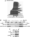
Fig. 1.
Fig. 1.
VBP1 specifically interacts with HIV-1 IN and mediates IN–pVHL interaction. (A) A yeast two-hybrid screening of a highly complex library of HIV-1 random fragment was performed by using VBP1 as bait. All obtained clones contained the IN gene, and their alignment allows the mapping of a VBP1 interacting domain in IN sequence located between residues 43 and 195. (B) HeLa cells or IN-HA cells (HeLa cells stably expressing IN-HA) were transfected with Myc-VBP1 or Myc-pVHL expression plasmids. Equal amounts of total cellular proteins (lysates) were immunoprecipitated by using anti-HA antibody. Immunoprecipitated proteins were then analyzed by Western blotting with anti-HA or anti-Myc antibody. (C) IN-HA cells were transfected with VBP1-specific (VBP1) or control luciferase (luc)-directed siRNA, prior to transfection with Myc-pVHL expression plasmid. After immunoprecipitation with anti-HA antibody, immunoprecipitates, as well as endogenous VBP1 and transfected Myc-pVHL in cell lysates, were analyzed by Western blotting by using anti-HA, anti-Myc, or anti-VBP1 antibodies, as indicated. Signals were quantified by densitometric analysis of the scanned autoradiographic films by using ImageJ software and revealed a 69% decrease of the coimmunoprecipitated Myc-pVHL/immunoprecipitated IN-HA ratio in the VBP1 siRNA-treated cells compared with the control cells.
Fig. 2.
Fig. 2.
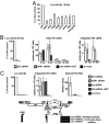
VBP1 and the Cul2/VHL ligase are important for HIV-1 gene expression at a postintegration step. (A) HeLa cells were transfected with siRNAs directed against indicated targets or control nontargeting (Ctrl) siRNA. Cells were subsequently infected with NL4-3ΔenvLuc VSVg virus, and luciferase activity was monitored in cell lysates 48 h after infection. (B) HeLa cells were transfected with either VBP1-directed (VBP1) or control nontargeting siRNA as indicated. Cells were subsequently infected with NL4-3ΔenvLuc VSVg virus or mock-infected, in the presence or absence of 3′-azido-3′-deoxythymidine, and luciferase activity was monitored in cell lysates. At different time points, DNA was extracted and subjected to real-time PCR analysis to quantify total viral DNA and integrated proviral DNA. (C) HeLa cells were transfected with either VBP1-directed, luciferase (luc)-directed, or control nontargeting siRNA as indicated. Cells were subsequently infected with NL4-3ΔenvLuc VSVg virus or mock-infected, in the presence or absence of 3′-azido-3′-deoxythymidine, and luciferase activity was monitored in cell lysates. Both DNA and RNA were extracted from each sample and subjected to real-time PCR or RT-PCR, respectively. Multiply spliced viral mRNAs (≈1.8 kb size class viral mRNAs) were selectively amplified by using a reverse primer encompassing the junction between the donor D4 and acceptor A7 splice sites involved in the formation of tat, rev, and nef mRNA. Integrated provirus and multiply spliced mRNAs were quantified for each sample. The scheme under the graphs represents the genetic organization of the HIV-1 genome and the ≈1.8-kb size class viral transcripts tat, rev, and nef (black bars) selectively amplified with the forward multiply spliced (FMS) and reverse multiply spliced (RMS) primers (arrows).
Fig. 3.
Fig. 3.
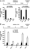
Integration is required before transcriptional control of HIV by VBP1. (A) HeLa cells were transfected with indicated siRNAs and subsequently infected with NL4-3ΔenvLuc VSVg virus (Left) or transfected with a plasmid encoding the NL4-3ΔenvLuc genome (Right). Luciferase activity was monitored in cell lysates 48 h after infection or transfection. (B) HeLa LTR-Luc cells were first transfected with indicated siRNAs and subsequently transfected with the indicated amount of a plasmid encoding Tat. Luciferase activity was monitored in cell lysates 48 h after transfection.
Fig. 4.
Fig. 4.
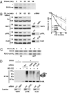
Prefoldin and Cul2/VHL complexes are involved in the ubiquitin–proteasome-dependent degradation of IN. (A) IN-HA cells were pulse-labeled with [35S]methionine/cysteine and subsequently chased for indicated time periods prior to immunoprecipitation by using anti-HA antibody. Immunoprecipitated proteins were analyzed by SDS/PAGE and fluorography. IN-HA and a stable contaminating protein (*) reflecting loading are visualized. Quantifications of two independent experiments resulted in an estimated IN half-life of 11 min. (B) IN-HA cells were transfected with siRNA specifically directed against VBP1 (VBP1a), pVHL, or Cul2 or with control luciferase (luc)-directed siRNA. Cells were subsequently treated with the protein synthesis inhibitor cycloheximide (100 μg/ml) for the indicated periods of time prior to lysis, and analyzed by Western blotting with anti-HA antibody and anti-α-tubulin antibody as an internal control. The effect of siRNAs on protein expression was monitored with specific antibodies. Chemiluminescence of the blots was acquired with a Fuji CCD camera (Kanagawa, Japan). For each condition, the IN-HA chemiluminescence signal was quantified by using Image Gauge software and normalized to the α-tubulin signal. Results from five to seven independent experiments are represented on the right. (C) pVHL-negative RCC4 cells stably transfected with pVHL (RCC4+pVHL) or not (RCC4) were transiently cotransfected with IN-HA and GFP expression plasmids. Cells were subsequently treated with cycloheximide (100 μg/ml) for the indicated times prior to lysis. Equal amounts of total protein lysates were then analyzed by Western blotting with anti-HA and anti-GFP antibodies. Chemiluminescence of the blots was quantified as in B, and the IN-HA signal was normalized to the GFP transfection control signal. (D) IN-HA or HeLa cells were transfected indicated siRNAs and subsequently treated with the proteasome inhibitor MG-132 (20 μM) or DMSO prior to lysis. Equal amounts of total cellular proteins were immunoprecipitated by using anti-HA antibody. Immunoprecipitated proteins were analyzed by Western blotting with anti-HA or anti-ubiquitin antibody (IP HA). VBP1 expression in cell lysates was monitored by anti-VBP1 immunoblotting (lysates).

Similar articles

Cited by

References

    1. Brown PO. Curr Top Microbiol Immunol. 1990;157:19–48. - PubMed
    1. Hehl EA, Joshi P, Kalpana GV, Prasad VR. J Virol. 2004;78:5056–5067. - PMC - PubMed
    1. Tasara T, Maga G, Hottiger MO, Hubscher U. FEBS Lett. 2001;507:39–44. - PubMed
    1. Tsurutani N, Kubo M, Maeda Y, Ohashi T, Yamamoto N, Kannagi M, Masuda T. J Virol. 2000;74:4795–4806. - PMC - PubMed
    1. Wu X, Liu H, Xiao H, Conway JA, Hehl E, Kalpana GV, Prasad V, Kappes JC. J Virol. 1999;73:2126–2135. - PMC - PubMed

Publication types