Urine proteomics: the present and future of measuring urinary protein components in disease
- PMID: 17698825
- PMCID: PMC1942114
- DOI: 10.1503/cmaj.061590
Urine proteomics: the present and future of measuring urinary protein components in disease
Abstract
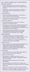
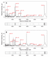
For centuries, physicians have attempted to use the urine for noninvasive assessment of disease. Today, urinalysis, in particular the measurement of proteinuria, underpins the routine assessment of patients with renal disease. More sophisticated methods for assessing specific urinary protein losses have emerged; however, albumin is still the principal urinary protein measured. Changes in the pattern of urinary protein excretion are not necessarily restricted to nephrourological disease; for instance, the appearance of beta-human chorionic gonadotropin in the urine of pregnant women is the basis for all commercially available pregnancy kits. Similarly, microalbuminuria is a clinically important marker not only of early diabetic nephropathy but also of concomitant cardiovascular disease. With the emergence of newer technologies, in particular mass spectrometry, it has become possible to study urinary protein excretion in even more detail. A variety of techniques have been used both to characterize the normal complement of urinary proteins and also to identify proteins and peptides that may facilitate earlier detection of disease, improve assessment of prognosis and allow closer monitoring of response to therapy. Such proteomics-based approaches hold great promise as the basis for new diagnostic tests and as the means to better understand disease pathogenesis. In this review, we summarize the currently available methods for urinary protein analysis and describe the newer approaches being taken to identify urinary biomarkers.
Figures


Similar articles
-
Proteomic analysis of urinary protein markers for accurate prediction of diabetic kidney disorder.J Assoc Physicians India. 2005 Jun;53:513-20. J Assoc Physicians India. 2005. PMID: 16121805
-
Urinary proteomics.Clin Chim Acta. 2007 Jan;375(1-2):49-56. doi: 10.1016/j.cca.2006.07.027. Epub 2006 Aug 1. Clin Chim Acta. 2007. PMID: 16942764 Review.
-
Pursuing type 1 diabetes mellitus and related complications through urinary proteomics.Transl Res. 2014 Mar;163(3):188-99. doi: 10.1016/j.trsl.2013.09.005. Epub 2013 Oct 3. Transl Res. 2014. PMID: 24096133
-
Identification of urinary protein pattern in type 1 diabetic adolescents with early diabetic nephropathy by a novel combined proteome analysis.J Diabetes Complications. 2005 Jul-Aug;19(4):223-32. doi: 10.1016/j.jdiacomp.2004.10.002. J Diabetes Complications. 2005. PMID: 15993357
-
Detection and measurement of urinary protein.Curr Opin Nephrol Hypertens. 2006 Nov;15(6):625-30. doi: 10.1097/01.mnh.0000247502.49044.10. Curr Opin Nephrol Hypertens. 2006. PMID: 17053478 Review.
Cited by
-
Collectives of diagnostic biomarkers identify high-risk subpopulations of hematuria patients: exploiting heterogeneity in large-scale biomarker data.BMC Med. 2013 Jan 17;11:12. doi: 10.1186/1741-7015-11-12. BMC Med. 2013. PMID: 23327460 Free PMC article.
-
Simplified microchip electrophoresis for rapid separation of urine proteins.J Clin Lab Anal. 2014 Mar;28(2):104-9. doi: 10.1002/jcla.21651. Epub 2014 Jan 6. J Clin Lab Anal. 2014. PMID: 24395581 Free PMC article.
-
Urinary pellet sample preparation for shotgun proteomic analysis of microbial infection and host-pathogen interactions.Methods Mol Biol. 2015;1295:65-74. doi: 10.1007/978-1-4939-2550-6_6. Methods Mol Biol. 2015. PMID: 25820714 Free PMC article.
-
Poor histological lesions in IgA nephropathy may be reflected in blood and urine peptide profiling.BMC Nephrol. 2013 Apr 11;14:82. doi: 10.1186/1471-2369-14-82. BMC Nephrol. 2013. PMID: 23577616 Free PMC article.
-
Urine Levels of Defensin α1 Reflect Kidney Injury in Leptospirosis Patients.Int J Mol Sci. 2016 Sep 27;17(10):1637. doi: 10.3390/ijms17101637. Int J Mol Sci. 2016. PMID: 27689992 Free PMC article.
References
-
- Viberti GC, Hill RD, Jarrett RJ, et al. Microalbuminuria as a predictor of clinical nephropathy in insulin-dependent diabetes mellitus. Lancet 1982;1:1430-2. - PubMed
-
- Abuelo JG. Proteinuria: diagnostic principles and procedures. Ann Intern Med 1983;98:186-91. - PubMed
-
- Free AH, Rupe CO, Metzler I. Studies with a new colorimetric test for proteinuria. Clin Chem 1957;3:716-27. - PubMed
-
- McElderry LA, Tarbit IF, Cassells-Smith AJ. Six methods for urinary protein compared. Clin Chem 1982;28:356-60. - PubMed
-
- Ginsberg JM, Chang BS, Matarese RA, et al. Use of single voided urine samples to estimate quantitative proteinuria. N Engl J Med 1983;309:1543-6. - PubMed
Publication types
MeSH terms
Substances
LinkOut - more resources
Full Text Sources
Other Literature Sources
