. 2007 Oct 5;282(40):29211-21.
doi: 10.1074/jbc.M610623200. Epub 2007 Aug 14.

A novel Ca(V)1.2 N terminus expressed in smooth muscle cells of resistance size arteries modifies channel regulation by auxiliary subunits

Affiliations

A novel Ca(V)1.2 N terminus expressed in smooth muscle cells of resistance size arteries modifies channel regulation by auxiliary subunits

Xiaoyang Cheng et al. J Biol Chem. .

Abstract

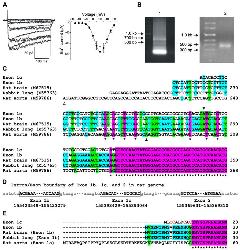
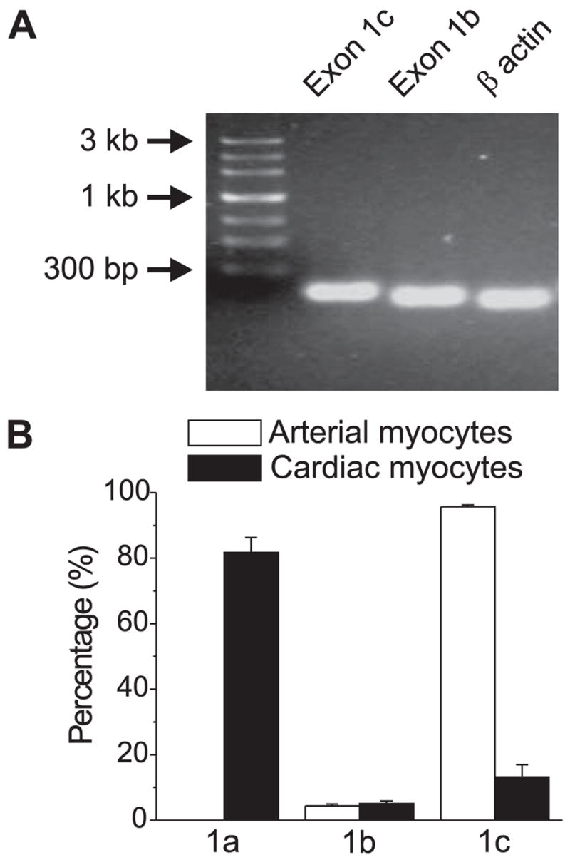
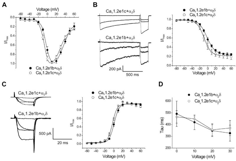
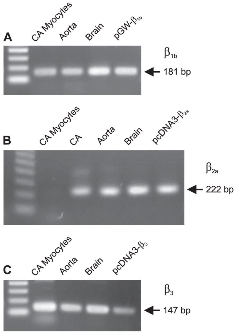
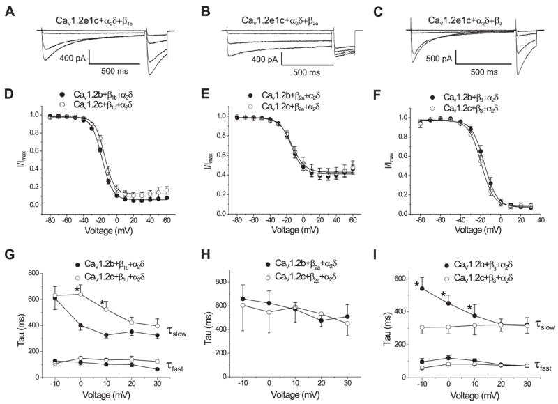
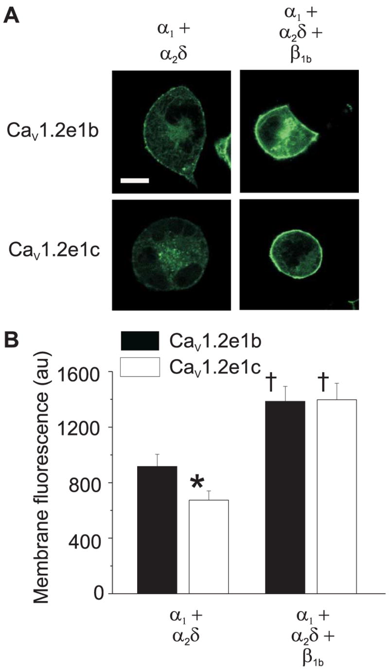
Voltage-dependent L-type Ca(2+) (Ca(V)1.2) channels are the principal Ca(2+) entry pathway in arterial myocytes. Ca(V)1.2 channels regulate multiple vascular functions and are implicated in the pathogenesis of human disease, including hypertension. However, the molecular identity of Ca(V)1.2 channels expressed in myocytes of myogenic arteries that regulate vascular pressure and blood flow is unknown. Here, we cloned Ca(V)1.2 subunits from resistance size cerebral arteries and demonstrate that myocytes contain a novel, cysteine rich N terminus that is derived from exon 1 (termed "exon 1c"), which is located within CACNA1C, the Ca(V)1.2 gene. Quantitative PCR revealed that exon 1c was predominant in arterial myocytes, but rare in cardiac myocytes, where exon 1a prevailed. When co-expressed with alpha(2)delta subunits, Ca(V)1.2 channels containing the novel exon 1c-derived N terminus exhibited: 1) smaller whole cell current density, 2) more negative voltages of half activation (V(1/2,act)) and half-inactivation (V(1/2,inact)), and 3) reduced plasma membrane insertion, when compared with channels containing exon 1b. beta(1b) and beta(2a) subunits caused negative shifts in the V(1/2,act) and V(1/2,inact) of exon 1b-containing Ca(V)1.2alpha(1)/alpha(2)delta currents that were larger than those in exon 1c-containing Ca(V)1.2alpha(1)/alpha(2)delta currents. In contrast, beta(3) similarly shifted V(1/2,act) and V(1/2,inact) of currents generated by exon 1b- and exon 1c-containing channels. beta subunits isoform-dependent differences in current inactivation rates were also detected between N-terminal variants. Data indicate that through novel alternative splicing at exon 1, the Ca(V)1.2 N terminus modifies regulation by auxiliary subunits. The novel exon 1c should generate distinct voltage-dependent Ca(2+) entry in arterial myocytes, resulting in tissue-specific Ca(2+) signaling.


Figures

FIGURE 1
FIGURE 1. Identification of the CaV1.2 exon 1c in rat cerebral arteries, relative location of exon 1c in the rat genome, and sequence comparison with exons 1a and 1b
A, voltage-dependent Ba2+ currents recorded in an isolated myocyte by depolarizing voltage steps (left panel) and a mean I-V relationship obtained from 10 cells (right panel). B, 5′-RACE products from cerebral arteries run in 0.8% agarose gel (left panel) with nested PCR products illustrating a band at ~500 bp (right panel). C, homology of exons 1a, 1b, and 1c compared with CaV1.2 nucleotide sequences from rat brain (M67515), rabbit lung (X55763), and rat aorta (M59786). The initiation sites of exon 1a, 1b, and 1c are indicated by △, ▲, and ▼, respectively. Part of exon 2 is also illustrated in purple. D, schematic diagram depicting the relative locations of exon 1b, 1c, and 2 within the rat genome. E, alignment of amino acid sequences encoded by CaV1.2 exon 1a, 1b, and 1c with part of the exon 2-encoded sequence shown in purple. * indicates identical nucleotides or amino acids.
FIGURE 2
FIGURE 2
A, RT-PCR revealed the presence of exons 1b and 1c in dissociated cerebral artery myocytes. β Actin is shown as a positive control. B, real-time PCR indicated the relative percentage of exons 1a, 1b, and 1c message in isolated rat cerebral artery myocytes and cardiac myocytes. Exon 1a was not detected in arterial myocytes.
FIGURE 3
FIGURE 3. Full-length CaV1.2 cDNAs cloned from rat small cerebral arteries
A, full-length CaV1.2e1b and CaV1.2e1c were cloned by RT-PCR and sub-cloned into pGEM-T Easy vector. Lane 1, CaV1.2e1b; lane 2, CaV1.2e1c; lane 3, CaV1.2e1c was released from pGEM-T easy vector by NotI digestion; lane 4, pGEM-T easy-CaV1.2e1c. B, CaV1.2e1b and CaV1.2e1c were subcloned into pIRES-hrGFP II vector, and the orientation direction of CaV1.2 in the expression vector was revealed by EcoRV digestion. Lane 1, pIRES-CaV1.2e1c-hrGFP II; lanes 2 and 3, pIRES-CaV1.2e1b-hrGFP II; lane 4, EcoRV digestion product of pIRES-CaV1.2e1b(+)-hrGFP II; and lane 5, EcoRV digestion product of pIRES-CaV1.2e1b(−)-hrGFP II.
FIGURE 4
FIGURE 4. Current-voltage (I-V) relationships of CaV1.2e1b and CaV1.2e1c channels when expressed with auxiliary subunits
Ba2+ currents were normalized to facilitate comparison. A, I-V relationships of CaV1.2e1b and CaV1.2e1c when co-expressed with α2δ. B, steady-state inactivation of CaV1.2e1c and CaV1.2e1b currents when co-expressed with α2δ. Exemplar current traces of 1-s conditioning depolarizing pulses evoked at −80, +10, and +30 mV followed by 200-ms test pulses to 0 mV (left panel). Cell capacitances for original recordings were: CaV1.2e1c + α2δ, 90 pF; CaV1.2e1b + α2δ, 66 pF. Mean steady-state inactivation fit with a Boltzmann function (right panel). C, voltage-dependent activation of CaV1.2e1c and CaV1.2e1b currents when co-expressed with α2δ. Exemplar tail currents evoked by repolarization to −80 mV after depolarizing test pulses to −20, 0, 20, and 40 mV (left panel). Cell capacitances for original recordings were: CaV1.2e1c + α2δ, 98 pF; CaV1.2e1b + α2δ, 66 pF. Mean voltage-dependent current activation (right panel). D, inactivation of CaV1.2e1b/α2δ and CaV1.2e1c/α2δ currents were best fit with a single exponential function. Tau was similar for CaV1.2e1b/α2δ and CaV1.2e1c/α2δ currents.
FIGURE 5
FIGURE 5. RT-PCR of CaVβ subunits expressed in dissociated rat cerebral arterial myocytes
A, β1b subunit. B, β2a subunit. C, β3 subunit. CA indicates cerebral artery.
FIGURE 6
FIGURE 6. Inactivation of CaV1.2e1b/α2δ and CaV1.2e1c/α2δ currents when expressed with β1b, β2a, or β3 subunits
A–C, original recordings of 1-s depolarizing pulses to −80, −20, −10, and 0 mV followed by 200-ms test pulses to 0 mV for each subunit combination indicated. D–F, voltage dependence of steady-state inactivation for each subunit combination. G–I, voltage dependence of inactivation constants for each combination specified. Inactivation of CaV1.2/α2δ/β1b and CaV1.2/α2δ/β3 currents were best fit with a bi-exponential function representing τfast and τslow, whereas CaV1.2/α2δ/β2a current inactivation was best fit with a single exponential. * illustrates p < 0.05.
FIGURE 7
FIGURE 7. Membrane localization of EGFP-tagged CaV1.2e1b and CaV1.2e1c channels when expressed with α2δ or with α2δ + β1b subunits in HEK 293 cells
A, representative images illustrating cellular fluorescence from EGFP-tagged CaV1.2e1b and CaV1.2e1c channels when expressed with subunit combinations indicated. Scale bar, 10 μm. B, mean data illustrating that co-expression of α2δ subunits elevated localized membrane fluorescence intensity of CaV1.2e1b more than for CaV1.2e1c, and that co-expression with α2δ + β1b subunits further increased membrane fluorescence and normalized the difference between CaV1.2e1b/α2δ and CaV1.2e1c/α2δ. au indicates arbitrary units. Number of cells: CaV1.2e1b/α2δ, 18; CaV1.2e1c/α2δ, 16; CaV1.2e1b/α2δ/β1b, 17; CaV1.2e1c/α2δ/β1b, 20. * indicates p < 0.05 when compared with CaV1.2e1b + α2δ, and †, p < 0.05 when compared with the same α1 subunit + α2δ.

References

    1. Gollasch M, Nelson MT. Kidney Blood Press Res. 2000;20:355–371. - PubMed
    1. Berridge MJ, Lipp P, Bootman MD. Nat Rev Mol Cell Biol. 2000;1:11–21. - PubMed
    1. Moosmang S, Schulla V, Welling A, Feil R, Feil S, Wegener JW, Hofmann F, Klugbauer N. EMBO J. 2003;22:6027–6034. - PMC - PubMed
    1. Nelson MT, Patlak JB, Worley JF, Standen NB. Am J Physiol. 1990;259:C3–18. - PubMed
    1. Sonkusare S, Palade PT, Marsh JD, Telemaque S, Pesic A, Rusch NJ. Vascul Pharmacol. 2006;44:131–142. - PMC - PubMed

Publication types

Substances

Associated data

LinkOut - more resources