Astrovirus increases epithelial barrier permeability independently of viral replication
- PMID: 17699569
- PMCID: PMC2168760
- DOI: 10.1128/JVI.00942-07
Astrovirus increases epithelial barrier permeability independently of viral replication
Abstract
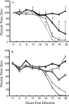
Astrovirus infection in a variety of species results in an age-dependent diarrhea; however, the means by which astroviruses cause diarrhea remain unknown. Studies of astrovirus-infected humans and turkeys have demonstrated few histological changes and little inflammation during infection, suggesting that intestinal damage or an overzealous immune response is not the primary mediator of astrovirus diarrhea. An alternative contributor to diarrhea is increased intestinal barrier permeability. Here, we demonstrate that astrovirus increases barrier permeability in a Caco-2 cell culture model system following apical infection. Increased permeability correlated with disruption of the tight-junction protein occludin and decreased the number of actin stress fibers in the absence of cell death. Additionally, permeability was increased when monolayers were treated with UV-inactivated virus or purified recombinant human astrovirus serotype 1 capsid in the form of virus-like particles. Together, these results demonstrate that astrovirus-induced permeability occurs independently of viral replication and is modulated by the capsid protein, a property apparently unique to astroviruses. Based on these data, we propose that the capsid contributes to diarrhea in vivo.
Figures


Similar articles
-
Oral Administration of Astrovirus Capsid Protein Is Sufficient To Induce Acute Diarrhea In Vivo.mBio. 2016 Nov 1;7(6):e01494-16. doi: 10.1128/mBio.01494-16. mBio. 2016. PMID: 27803180 Free PMC article.
-
Type I Interferon Response Limits Astrovirus Replication and Protects against Increased Barrier Permeability In Vitro and In Vivo.J Virol. 2015 Dec 9;90(4):1988-96. doi: 10.1128/JVI.02367-15. Print 2016 Feb 15. J Virol. 2015. PMID: 26656701 Free PMC article.
-
Physiologically relevant increase in temperature causes an increase in intestinal epithelial tight junction permeability.Am J Physiol Gastrointest Liver Physiol. 2006 Feb;290(2):G204-12. doi: 10.1152/ajpgi.00401.2005. Am J Physiol Gastrointest Liver Physiol. 2006. PMID: 16407590
-
Astrovirus Biology and Pathogenesis.Annu Rev Virol. 2017 Sep 29;4(1):327-348. doi: 10.1146/annurev-virology-101416-041742. Epub 2017 Jul 17. Annu Rev Virol. 2017. PMID: 28715976 Review.
-
Pathogenesis of astrovirus infection.Viral Immunol. 2005;18(1):4-10. doi: 10.1089/vim.2005.18.4. Viral Immunol. 2005. PMID: 15802949 Review.
Cited by
-
Impact of adenoviral stool load on adenoviremia in pediatric hematopoietic stem cell transplant recipients.Pediatr Infect Dis J. 2015 Jun;34(6):562-5. doi: 10.1097/INF.0000000000000678. Pediatr Infect Dis J. 2015. PMID: 25742243 Free PMC article.
-
Human immune deficiency virus conundrum: An everlasting challenge!Oman J Ophthalmol. 2022 Mar 2;15(1):13-19. doi: 10.4103/ojo.ojo_125_21. eCollection 2022 Jan-Apr. Oman J Ophthalmol. 2022. PMID: 35388268 Free PMC article.
-
Astrovirus replication in human intestinal enteroids reveals multi-cellular tropism and an intricate host innate immune landscape.PLoS Pathog. 2019 Oct 31;15(10):e1008057. doi: 10.1371/journal.ppat.1008057. eCollection 2019 Oct. PLoS Pathog. 2019. PMID: 31671153 Free PMC article.
-
Gualou Xiebai Decoction ameliorates increased Caco-2 monolayer permeability induced by bile acids via tight junction regulation, oxidative stress suppression and apoptosis reduction.J Bioenerg Biomembr. 2022 Feb;54(1):45-57. doi: 10.1007/s10863-021-09927-y. Epub 2021 Oct 31. J Bioenerg Biomembr. 2022. PMID: 34718922
-
Suppression of astrovirus replication by an ERK1/2 inhibitor.J Virol. 2008 Aug;82(15):7475-82. doi: 10.1128/JVI.02193-07. Epub 2008 May 28. J Virol. 2008. PMID: 18508903 Free PMC article.
References
-
- Belliot, G., H. Laveran, and S. S. Monroe. 1997. Detection and genetic differentiation of human astroviruses: phylogenetic grouping varies by coding region. Arch. Virol. 142:1323-1334. - PubMed
-
- Chapron, C. D., N. A. Ballester, J. H. Fontaine, C. N. Frades, and A. B. Margolin. 2000. Detection of astroviruses, enteroviruses, and adenovirus types 40 and 41 in surface waters collected and evaluated by the information collection rule and an integrated cell culture-nested PCR procedure. Appl. Environ. Microbiol. 66:2520-2525. - PMC - PubMed
Publication types
MeSH terms
Substances
Grants and funding
LinkOut - more resources
Full Text Sources
Miscellaneous

