. 2007 Nov;81(21):11577-84.
doi: 10.1128/JVI.01414-07. Epub 2007 Aug 15.

The topology of hepatitis B virus pregenomic RNA promotes its replication

Affiliations

The topology of hepatitis B virus pregenomic RNA promotes its replication

Teresa M Abraham et al. J Virol. 2007 Nov.

Abstract

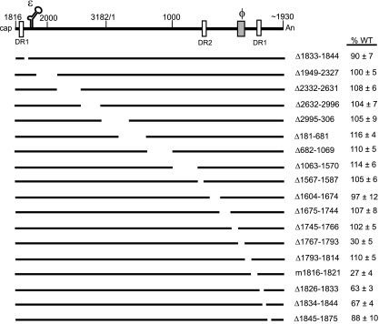
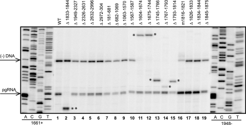
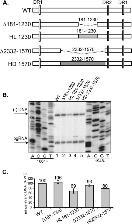
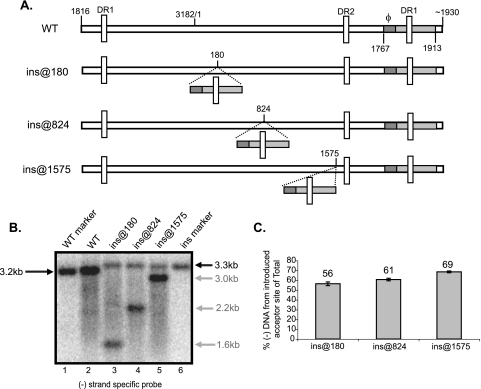
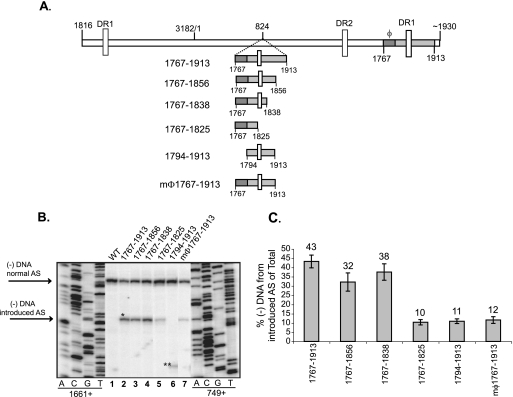
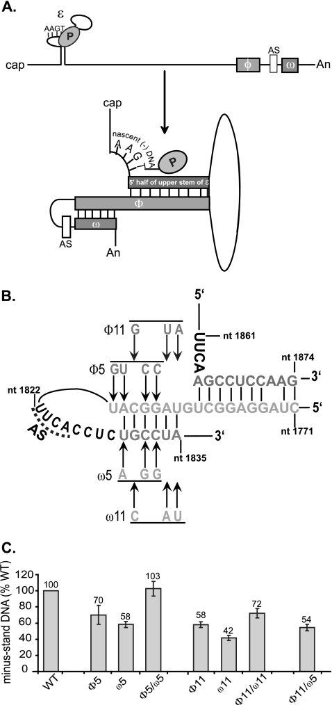
Previous analysis of hepatitis B virus (HBV) indicated base pairing between two cis-acting sequences, the 5' half of the upper stem of epsilon and phi, contributes to the synthesis of minus-strand DNA. Our goal was to identify other cis-acting sequences on the pregenomic RNA (pgRNA) involved in the synthesis of minus-strand DNA. We found that large portions of the pgRNA could be deleted or substituted without an appreciable decrease in the level of minus-strand DNA synthesized, indicating that most of the pgRNA is dispensable and that a specific size of the pgRNA is not required for this process. Our results indicated that the cis-acting sequences for the synthesis of minus-strand DNA are present near the 5' and 3' ends of the pgRNA. In addition, we found that the first-strand template switch could be directed to a new location when a 72-nucleotide (nt) fragment, which contained the cis-acting sequences present near the 3' end of the pgRNA, was introduced at that location. Within this 72-nt region, we uncovered two new cis-acting sequences, which flank the acceptor site. We show that one of these sequences, named omega and located 3' of the acceptor site, base pairs with phi to contribute to the synthesis of minus-strand DNA. Thus, base pairing between three cis-acting elements (5' half of the upper stem of epsilon, phi, and omega) are necessary for the synthesis of HBV minus-strand DNA. We propose that this topology of pgRNA facilitates first-strand template switch and/or the initiation of synthesis of minus-strand DNA.


Figures

FIG. 1.
FIG. 1.
Schematic representation of HBV WT pgRNA and variants. The black lines represent either full-length pgRNA or pgRNAs expressed from variants. The white vertical boxes represent the direct repeat (DR) sequences, DR1:1822-1832 and DR2:1588-1598. The 5′ end of pgRNA is at nt 1816. The poly(A) tail begins at approximately nt 1930. The encapsidation signal, ɛ (nt 1845 to 1905), and a previously identified cis element, φ (nt 1771 to 1789), are indicated. The names of the deletion variants reflect the deleted nucleotides on the HBV pgRNA (inclusive). The m1816-1821 variant changes 6 nt to the respective Watson-Crick partners. On the right are the mean values and standard deviations of the relative levels of minus-strand DNA compared to the WT reference. The ability of a virus to synthesize minus-strand DNA was determined as the level of minus-strand DNA divided by the sum of pgRNA and minus-strand DNA levels. The mean values represent analysis from at least six independent transfections of each variant. The representations of the deleted sequences are not drawn to scale.
FIG. 2.
FIG. 2.
Representative primer extension analysis of variants. Replicative intermediates of WT reference and variants were isolated from cytoplasmic capsids from Huh7 cells and analyzed by primer extension to measure the levels of pgRNA and minus-strand DNA. The oligonucleotides 1661+ and 1948 were used. The positions of the 5′ ends of pgRNA and minus-strand DNA are indicated by arrows on the left. The single asterisk indicates the altered positions at which minus-strand DNA migrates reflecting the size of the deletion. The double asterisks indicate the altered position at which pgRNA migrates, reflecting the size of the deletion. Oligonucleotide 1556+ was used instead of 1661+ to measure levels of minus-strand DNA for Δ1604-1674 and Δ1675-1744 (lanes 11 and 12).
FIG. 3.
FIG. 3.
Large deletions and heterologous substitutions of the pgRNA are tolerated during the synthesis of minus-strand DNA. (A) WT HBV and HBV/LacZ (HL) or HBV/DHBV (HD) chimeric variants. The white rectangles represent HBV sequences. The dotted lines represent deletions. The coordinates of the deletions are inclusive. The light and dark gray boxes represent LacZ and DHBV sequences, respectively, that were substituted for HBV sequences. The vertical white rectangles represent the DR sequences. (B) Primer extension analysis of replicative intermediates of WT and deletion and substitution variants harvested from cytoplasmic capsids from Huh7 cells. Oligonucleotides 1661+ and 1948 were used to measure minus-strand DNA and pgRNA, respectively. The 5′ ends of minus-strand DNA and pgRNA are indicated on the left. (C) Proportions of minus-strand DNA relative to the WT reference. The level of synthesis of minus-strand DNA is defined as the amount of minus-strand DNA divided by the sum of pgRNA and minus-strand DNA. The values for all variants are normalized to the WT reference. The mean values are from at least six independent transfections of each variant. The error bars indicate standard deviations.
FIG. 4.
FIG. 4.
Nucleotides 1767 to 1913 direct the first-strand template switch to a new AS when introduced at different locations on the pgRNA. (A) Representation of WT HBV and insertion variants. The gray rectangle represents nt 1767 to 1913 (inclusive) and contains φ (dark gray). For variants ins@180, ins@824, and ins@1575, nt 1767 to 1913 have been inserted after nt 180, 824, and 1575, respectively. The location of the direct repeat (DR) sequences are indicated by vertical white rectangles. The nucleotide coordinates of the 5′ and 3′ ends of pgRNA are indicated. (B) Southern blot of WT and insertion variants. DNA replicative intermediates harvested from Huh7 cells were denatured prior to electrophoresis. A minus-strand specific RNA probe spanning nt 3096 to 264 was used to detect minus-strand DNA. This probe does not detect DNA synthesized from encapsidated spliced RNA. WT and insertion genome length markers were used to indicate the size of single-stranded DNA arising from the AS (lanes 1 and 6). The sizes of minus-strand DNA arising from normal and introduced AS are indicated in black and gray, respectively. (C) For each insertion variant, gray bars represent minus-strand DNA arising from introduced AS as a proportion of total minus-strand DNA. Total minus-strand DNA is the sum of completely elongated minus-strand DNA from normal and introduced AS. Minus-strand DNA derived from spliced RNA are not detected in this analysis due to probe choice. The mean values represent analysis from at least six independent transfections of each variant. The error bars indicate standard deviations.
FIG. 5.
FIG. 5.
Nucleotides 1767 to 1838 are sufficient to direct first-strand template switch to a new AS when introduced at another location on the pgRNA. (A) WT HBV and insertion variants. The gray rectangle represents the sequence introduced after nt 824. The dark gray rectangle represents φ. The 5′ and 3′ ends of the introduced sequence are indicated. The coordinates are inclusive of region inserted after nt 824. The locations of the DR sequences are indicated by vertical white rectangles. The 5′ and 3′ ends of the pgRNA are also indicated. (B) Primer extension analysis of WT and insertion variants. Oligonucleotides 1661+ and 749+ were used to measure minus-strand DNA arising from the normal and introduced AS, respectively. The 5′ ends of minus-strand DNA arising from normal and introduced AS are indicated. The single and double asterisks indicate the positions of minus-strand DNA arising from the introduced AS. The double asterisks indicate the altered position of minus-strand DNA reflecting the 27-nt difference between the oligonucleotide 749+ annealing site and the introduced AS. (C) For each insertion variant, the gray bars represent minus-strand DNA arising from the introduced AS as a proportion of total minus-strand DNA. Total minus-strand DNA is determined as the sum of minus-strand DNA from the normal and introduced AS. Primer extension detects all minus-strand DNAs within the capsid, i.e., incompletely elongated minus-strand DNAs and minus-strand DNA derived from pgRNA and spliced RNAs. This could account for the difference in levels of minus-strand DNA from the introduced AS for ins@824 in the analysis in Fig. 4C. The mean values represent analysis from at least six independent transfections. The error bars represent the standard deviation.
FIG. 6.
FIG. 6.
Base pairing between ω and a portion of φ contributes to the synthesis of minus-strand DNA. (A) Schematic representation of the dynamic conformation of pgRNA. The 5′ half of upper stem of ɛ, φ, and ω are shown as gray rectangles. The nascent minus-strand DNA attached to P protein is shown on the bulge of ɛ. The 4-nt AS upstream of ω is shown as a white rectangle. The figure is not drawn to scale. (B) Putative base pairing between the 5′ half of the upper stem of ɛ, φ, and ω. Substitutions designed to disrupt base pairing between φ and ω are shown. Variants that restore base pairing combine the corresponding φ and ω substitutions. P, P protein; AS, AS for minus-strand DNA synthesis. (C) Proportion of minus-strand DNA compared to WT reference. The level of synthesis of minus-strand DNA is defined as the amount of minus-strand DNA divided by the sum of pgRNA and minus-strand DNA. The mean values represent analysis from at least six independent transfections of each variant. The error bars represent the standard deviation.

Similar articles

Cited by

References

    1. Abraham, T. M., and D. D. Loeb. 2006. Base Pairing between the 5′ half of ɛ and a cis-acting sequence, φ, makes a contribution to the synthesis of minus-strand DNA for human hepatitis B virus. J. Virol. 80:4380-4387. - PMC - PubMed
    1. Bartenschlager, R., and H. Schaller. 1992. Hepadnaviral assembly is initiated by polymerase binding to the encapsidation signal in the viral RNA genome. EMBO J. 11:3413-3420. - PMC - PubMed
    1. Beck, J., and M. Nassal. 1998. Formation of a functional hepatitis B virus replication initiation complex involves a major structural alteration in the RNA template. Mol. Cell. Biol. 18:6265-6272. - PMC - PubMed
    1. Fallows, D., and S. Goff. 1995. Mutations in the epsilon sequences of human hepatitis B virus affect both RNA encapsidation and reverse transcription. J. Virol. 69:3067-3073. - PMC - PubMed
    1. Geballe, A. P., R. R. Spaete, and E. S. Mocarski. 1986. A cis-acting element within the 5′ leader of a cytomegalovirus β transcript determines kinetic class. Cell 46:865-872. - PubMed

Publication types