. 2007 Nov;81(21):11850-60.
doi: 10.1128/JVI.01421-07. Epub 2007 Aug 15.

Protein kinase CK2 phosphorylation of EB2 regulates its function in the production of Epstein-Barr virus infectious viral particles

Affiliations

Protein kinase CK2 phosphorylation of EB2 regulates its function in the production of Epstein-Barr virus infectious viral particles

Cahora Medina-Palazon et al. J Virol. 2007 Nov.

Abstract

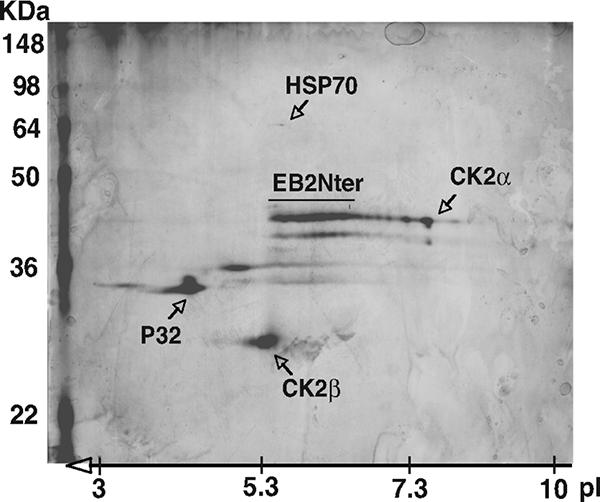
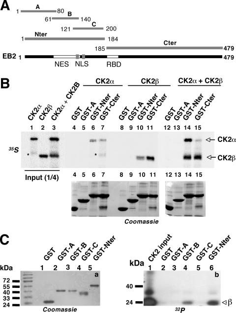
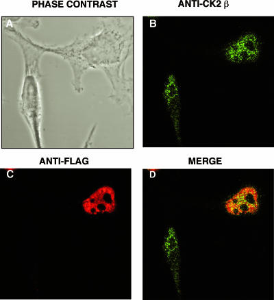
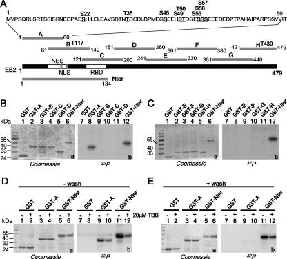
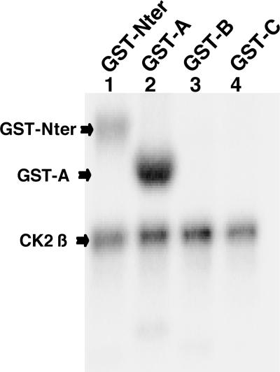
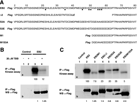
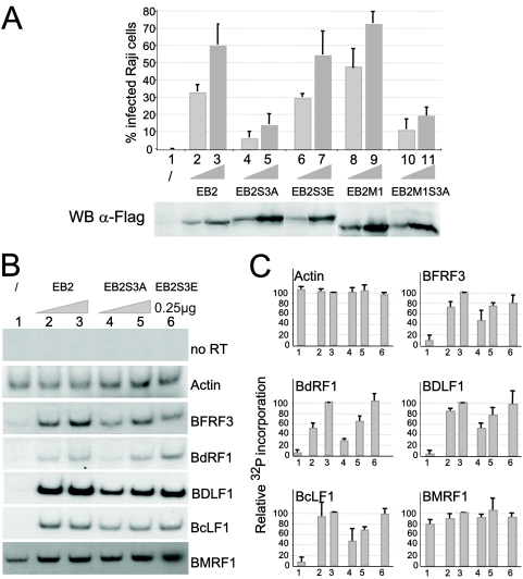
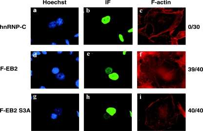
The Epstein-Barr Virus (EBV) early protein EB2 (also called BMLF1, Mta, or SM) promotes the nuclear export of a subset of early and late viral mRNAs and is essential for the production of infectious virions. We show here that in vitro, protein kinase CK2alpha and -beta subunits bind both individually and, more efficiently, as a complex to the EB2 N terminus and that the CK2beta regulatory subunit also interacts with the EB2 C terminus. Immunoprecipitated EB2 has CK2 activity that phosphorylates several sites within the 80 N-terminal amino acids of EB2, including Ser-55, -56, and -57, which are localized next to the nuclear export signal. EB2S3E, the phosphorylation-mimicking mutant of EB2 at these three serines, but not the phosphorylation ablation mutant EB2S3A, efficiently rescued the production of infectious EBV particles by HEK293(BMLF1-KO) cells harboring an EB2-defective EBV genome. The defect of EB2S3A in transcomplementing 293(BMLF1-KO) cells was not due to impaired nucleocytoplasmic shuttling of the mutated protein but was associated with a decrease in the cytoplasmic accumulation of several late viral mRNAs. Thus, EB2-mediated production of infectious EBV virions is regulated by CK2 phosphorylation at one or more of the serine residues Ser-55, -56, and -57.


Figures

FIG. 1.
FIG. 1.
Both CK2α and -β subunits copurify with the 184 EB2 N-terminal amino acids. HEK293T cells were transfected with pNTAP.EB2Nter, expressing the EB2Nter protein fused at the N terminus to a calmodulin-binding peptide and to a streptavidin-binding peptide. TAP-tagged EB2 and associated proteins expressed in HEK293T cells were sequentially purified on streptavidin and calmodulin columns. The purified proteins were separated by 2D gel, silver stained, and identified by MALDI-TOF mass spectrometry. The unlabeled spots were not identified by mass spectrometry. The positions of the molecular mass markers are indicated on the left of the gel. pI, isoelectric point.
FIG. 2.
FIG. 2.
CK2 binding to EB2. (A) Schematic representation of EB2 and EB2 polypeptides used to generate GST fusion proteins. NLS, nuclear localization signal; RBD, RNA-binding domain. (B) Recombinant CK2α and CK2β in vitro labeled with [35S]methionine (lanes 1 to 3), either individually (lanes 4 to 11) or mixed together (lanes 12 to 15), for 15 min at 4°C were incubated with glutathione-Sepharose 4B beads loaded with GST or GST-A, -Nter, or -Cter. Proteins bound to the beads were eluted, resolved by SDS-PAGE, and visualized by Coomassie blue staining (bottom, lanes 4 to 15) and by autoradiography (top, lanes 4 to 15). *, nonspecific band. (C) Purified CK2, 32P labeled by autophosphorylation in vitro (b, lane 1), was incubated with beads loaded with GST or GST-A, -B, -C, and -Nter. Proteins bound to the beads were eluted, resolved by SDS-PAGE, and visualized by Coomassie blue staining (a) and by autoradiography (b).
FIG. 3.
FIG. 3.
EB2 and CK2β colocalize in the nuclei of living cells. HeLa cells expressing F.EB2 were immunostained using a rabbit anti-CK2β antibody and an Alexa Fluor 488-conjugated secondary antibody (B) and the M2 anti-Flag MAb and a Fluorolink Cy3-conjugated secondary antibody (C). (D) Colocalization of CK2β and EB2. The cell fluorescence was examined using confocal microscopy. Panel A shows the same cells in phase-contrast microscopy.
FIG. 4.
FIG. 4.
CK2 in HEK293T cell extracts binds to and phosphorylates the EB2 N terminus. (A) Schematic representation of the EB2 peptides fused to GST to generate GST-A, -B, -C, -D, -E, -F, -G, -H, and -Nter. Putative CK2 sites shown over the sequences of peptides A, B, and H were determined using Scansite (http://scansite.mit.edu). (B and C) Glutathione-Sepharose 4B beads loaded with GST and the GST fusion proteins indicated above the lanes were incubated for 30 min at 30°C with a HEK293T cell extract supplemented with kinase buffer and [γ-32P]ATP. GST and the GST fusion proteins were eluted, resolved by SDS-PAGE, and visualized by Coomassie blue staining (gels a) and by autoradiography (gels b). (D and E) Glutathione-Sepharose 4B beads loaded with GST and the GST fusion proteins indicated above the lanes were incubated overnight with a HEK293T cell extract. The − wash assays were supplemented with kinase buffer and [γ-32P]ATP and further incubated for 30 min at 30°C. For the + wash assays, the beads were washed extensively with MTPBS and then submitted to a kinase assay. Where indicated, 20 μM TBB was added to the assay. GST and GST fusion proteins were eluted, resolved by SDS-PAGE, and visualized by Coomassie blue staining (gels a, lanes 1 to 6) and by autoradiography (gels b, lanes 7 to 12).
FIG. 5.
FIG. 5.
Purified CK2 holoenzyme phosphorylation sites are restricted to the N-terminal 80 amino acids of EB2. Glutathione-Sepharose 4B beads loaded with GST-Nter, -A, -B, and -C were incubated with purified CK2 holoenzyme in an in vitro kinase assay. The GST fusion proteins were eluted, resolved by SDS-PAGE, and analyzed by autoradiography.
FIG. 6.
FIG. 6.
EB2 immunoprecipitates contains catalytically active CK2. (A) Amino acid sequences of the EB2 N-terminal 80 amino acids and of EB2 mutants Δ2, S3A, S3E, M1, and M1S3A. The Flag tag is in bold and italics. Amino acids added during the cloning procedure in Δ2 are in bold and italics. Putative CK2 sites are shown by a dot under the EB2 sequence. (B) Extracts from HEK293T cells, mock transfected or expressing F.EB2, were incubated with the M2 anti-Flag affinity gel. Immune complexes were submitted to an in vitro kinase assay in the absence (lanes 1 and 3) or presence (lanes 2 and 4) of the CK2-specific inhibitor TBB (20 μM). Proteins in the immune complexes were resolved by SDS-PAGE and visualized by Western blotting (b) or autoradiography (a). IP, immunoprecipitation. (C) F.EB2, F.EB2M1, F.EB2Δ2, F.EB2S3A, F.EB2S3E, and F.EB2M1S3A expressed in HEK293T cells were analyzed as for panel B. In panels B and C, the relative amounts of EB2 immunoprecipitated were quantified using the Odyssey infrared imaging system. The relative intensities of the radioactive bands on the membrane were quantified using a PhosphorImager. The numbers under blots a correspond to the intensities of the radioactive bands (expressed in arbitrary units compared to EB2) normalized against the amounts of proteins immunoprecipitated (expressed in arbitrary units by comparison to EB2, shown by the numbers under blots b).
FIG. 7.
FIG. 7.
The EB2S3A mutant does not rescue infectious-virus production by 293BMLF1-KO cells and inefficiently exports several late EBV mRNAs. (A) 293BMLF1-KO cells were transfected with 1 μg of EB1 expression vector alone (lane 1) or together with 0.25 μg (lanes 2, 4, 6, 8, and 10) or with 0.5 μg (lanes 3, 5, 7, 9, and 11) of F.EB2, F.EB2S3A, F.EB2S3E, F.EB2M1, and F.EB2M1S3A expression vectors. Three days after transfection, Raji cells were infected with the filtered medium, and after 48 h, infected cells were quantified by FACS analysis (FACSCALIBUR). Two representative Raji cell infections are presented. All errors bars represent standard deviations. WB anti-FLAG, levels of expression of F.EB2 and F.EB2 mutated proteins in transfected 293BMLF1-KO cells, as evaluated by Western blotting with the M2 anti-FLAG MAb. (B) Cytoplasmic RNAs were extracted from 293BMLF1-KO cells transfected as described for panel A, except that RNA was extracted only from cells transfected with 0.25 μg of F.EB2S3E (lane 6). They were reverse transcribed using oligo(dT)18 as a primer and PCR amplified in the presence of 0.1 μCi [α-32P]dCTP with the specific primers shown in Table 1. The DNA fragments generated by PCR were separated on a 6% polyacrylamide gel and revealed by autoradiography. (C) The relative amounts of 32P incorporated in the PCR products presented in panel B were quantified using a PhosphorImager (Amersham). The results are presented as 32P incorporation relative to that obtained at the highest EB2 concentration, arbitrarily expressed as 100 (lanes 3). All errors bars represent standard deviations.
FIG. 8.
FIG. 8.
Nucleocytoplasmic shuttling of EB2 is not affected by the S3A mutation. F.EB2, and F.EB2S3A were expressed in HeLa cells. After 48 h, the HeLa cells were cocultivated overnight with NIH 3T3 cells. Prior to PEG-induced cell fusion, the cells were incubated for 2 h with cycloheximide. After fusion, the cells were further incubated for 2 h with cycloheximide. The cells were then immunostained using either the M2 anti-Flag MAb (panels e and h) or the anti-hnRNP-C (4F4) MAb (panel b) and an Alexa Fluor-conjugated secondary antibody. F-actin was labeled with Alexa Fluor 546 phalloidin to visualize the heterokaryons (panels c, f, and i). The cell nucleus was stained with Hoescht dye (panels a, d, and g). On the right sides of the panels the numbers indicate the numbers of heterokaryons with positive mouse cell nuclei immunostaining (indicating protein shuttling)/the total number of heterokaryons with positive HeLa nuclei immunostaining. IF, immunofluorescence.

References

    1. Aguilera, A. 2005. Cotranscriptional mRNP assembly: from the DNA to the nuclear pore. Curr. Opin. Cell Biol. 17:242-250. - PubMed
    1. Batisse, J., E. Manet, J. Middeldorp, A. Sergeant, and H. Gruffat. 2005. The Epstein-Barr virus mRNA export factor EB2 is essential for intranuclear capsid assembly and production of gp350. J. Virol. 79:14102-14111. - PMC - PubMed
    1. Bryant, H. E., D. A. Matthews, S. Wadd, J. E. Scott, J. Kean, S. Graham, W. C. Russell, and B. Clements. 2000. Interaction between herpes simplex virus type 1 IE63 protein and cellular protein p32. J. Virol. 74:11322-11328. - PMC - PubMed
    1. Buisson, M., F. Hans, I. Kusters, N. Duran, and A. Sergeant. 1999. The C-terminal region but not the Arg-X-Pro repeat of Epstein-Barr virus protein EB2 is required for its effect on RNA splicing and transport. J. Virol. 73:4090-4100. - PMC - PubMed
    1. Chen, I. B., L. Li, L. Silva, and R. M. Sandri-Goldin. 2005. ICP27 recruits Aly/REF but not TAP/NXF1 to herpes simplex virus type 1 transcription sites although TAP/NXF1 is required for ICP27 export. J. Virol. 79:3949-3961. - PMC - PubMed

Publication types