. 2007 Oct;81(20):11372-80.
doi: 10.1128/JVI.01327-07. Epub 2007 Aug 15.

Novel adeno-associated virus serotypes efficiently transduce murine photoreceptors

Affiliations

Novel adeno-associated virus serotypes efficiently transduce murine photoreceptors

Mariacarmela Allocca et al. J Virol. 2007 Oct.

Abstract

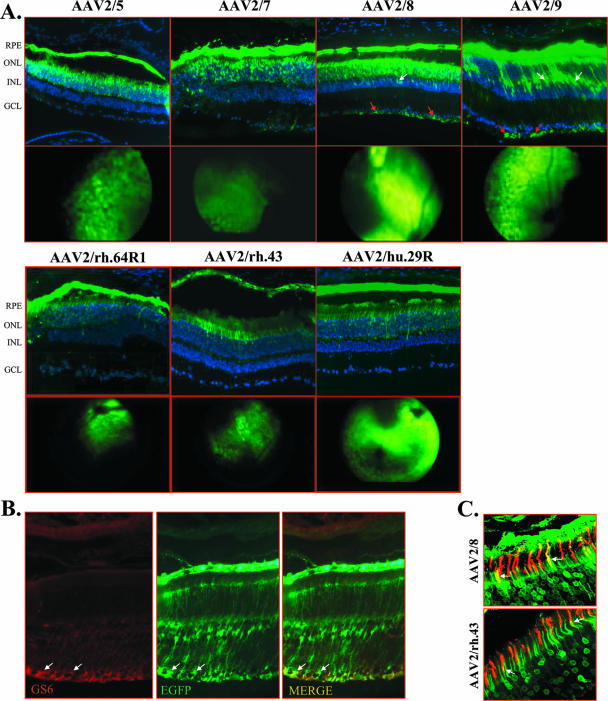
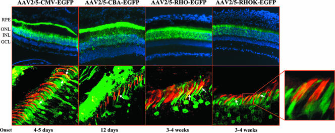
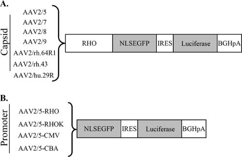
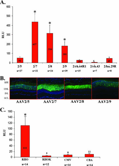
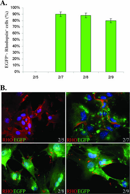
Severe inherited retinal diseases, such as retinitis pigmentosa and Leber congenital amaurosis, are caused by mutations in genes preferentially expressed in photoreceptors. While adeno-associated virus (AAV)-mediated gene transfer can correct retinal pigment epithelium (RPE) defects in animal models, approaches for the correction of photoreceptor-specific diseases are less efficient. We evaluated the ability of novel AAV serotypes (AAV2/7, AAV2/8, AAV2/9, AAV2rh.43, AAV2rh.64R1, and AAV2hu.29R) in combination with constitutive or photoreceptor-specific promoters to improve photoreceptor transduction, a limiting step in photoreceptor rescue. Based on a qualitative analysis, all AAV serotypes tested efficiently transduce the RPE as well as rod and cone photoreceptors after subretinal administration in mice. Interestingly, AAV2/9 efficiently transduces Müller cells. To compare photoreceptor transduction from different AAVs and promoters in both a qualitative and quantitative manner, we designed a strategy based on the use of a bicistronic construct expressing both enhanced green fluorescent protein and luciferase. We found that AAV2/8 and AAV2/7 mediate six- to eightfold higher levels of in vivo photoreceptor transduction than AAV2/5, considered so far the most efficient AAV serotype for photoreceptor targeting. In addition, following subretinal administration of AAV, the rhodopsin promoter allows significantly higher levels of photoreceptor expression than the other ubiquitous or photoreceptor-specific promoters tested. Finally, we show that AAV2/7, AAV2/8, and AAV2/9 outperform AAV2/5 following ex vivo transduction of retinal progenitor cells differentiated into photoreceptors. We conclude that AAV2/7 or AAV2/8 and the rhodopsin promoter provide the highest levels of photoreceptor transduction both in and ex vivo and that this may overcome the limitation to therapeutic success observed so far in models of inherited severe photoreceptor diseases.


Figures

FIG. 1.
FIG. 1.
Subretinal administration of novel AAV serotypes expressing EGFP in adult C57BL/6 mice. (A) The first and third rows depict fluorescence microscopy evaluations of EGFP expression 4 weeks after subretinal injection with AAV2/5-CMV-EGFP, AAV2/7-CMV-EGFP, AAV2/8-CMV-EGFP, AAV2/9-CMV-EGFP, AAV2/rh.64R1-CMV-EGFP, AAV2/rh.43-CMV-EGFP, and AAV2/rh.29R-CMV-EGFP. Abbreviations: ONL, outer nuclear layer; INL, inner nuclear layer; GCL, ganglion cell layer. White arrows, Müller cell nuclei; red arrows, Müller cell endfoot membranes. The second and fourth rows depict in vivo imaging of EGFP fluorescence in mouse retinas 5 days after subretinal injections of AAV2/5-CMV-EGFP, AAV2/7-CMV-EGFP, and AAV2/8-CMV-EGFP, 11 days after injection of AAV2/9-CMV-EGFP, and 7 days after injection of AAV2/rh.64R1-CMV-EGFP, AAV2/rh.43-CMV-EGFP, and AAV2/rh.29R-CMV-EGFP. Fundus photographs show punctate EGFP fluorescence diffusing from the site of the subretinal injection to 30 to 40% of the treated retina. (B) Müller cell transduction by AAV2/9 vectors expressing EGFP from the CMV promoter. Staining with anti-GS6 antibody (GS6; red label) on retinal sections of animals injected with AAV2/9-CMV-EGFP is shown. Colocalization of EGFP expression and GS6 staining is indicated by the arrows (MERGE). Microscope magnification, ×20. (C) Cone transduction from novel AAV serotypes expressing EGFP from the CMV promoter. Staining with PNA-lectin (red label) on retinal sections of animals injected with AAV2/8-CMV-EGFP or AAV2/rh.43-CMV-EGFP is shown. Colocalization of EGFP expression and PNA staining is indicated by the arrows. Confocal microscope magnification, ×63.
FIG. 2.
FIG. 2.
Subretinal injections of AAV2/5-EGFP vectors with various promoters into adult C57BL/6 mice. The images in the top row depict fluorescence microscopy evaluation of EGFP expression 4 weeks after subretinal injections of AAV2/5 encoding EGFP from the CMV, CBA, RHO, and RHOK promoters. Abbreviations: ONL, outer nuclear layer; INL, inner nuclear layer; GCL, ganglion cell layer. The images in the bottom row show staining with PNA-lectin (red label) on retinal sections of animals injected with AAV2/5-CMV-EGFP, AAV2/5-CBA-EGFP, AAV2/5-RHO-EGFP, and AAV2/5-RHOK-EGFP. Colocalization of EGFP expression and PNA staining is indicated by the arrows. Confocal microscope magnification, ×63. The selected field shows the colocalization of cone sheathes (PNA, red label) and EGFP expression (green label) at increased magnification. The magnified portion depicts the onset of gene expression as assessed by indirect ophthalmoscopy.
FIG. 3.
FIG. 3.
Strategy to assess levels of transgene expression following AAV-mediated photoreceptor gene transfer. (A) To identify the AAV serotype that results in the highest number of transduced photoreceptors, a bicistronic reporter was expressed in photoreceptors using the photoreceptor-specific RHO promoter. The bicistronic reporter includes the sequence of NLSEGFP (see Results and Discussion for details) followed by that of luciferase. NLSEGFP allows the qualitative analysis of photoreceptor transduction by histology, while luciferase allows us to quantify the transduction levels by a luminometric assay. (B) To identify the promoter that results in the highest levels of expression in photoreceptors, we generated AAV2/5 vectors encoding the bicistronic reporter under the transcriptional control of various promoters. IRES, internal ribosomal entry site sequence; BGHpA, bovine growth hormone poly(A).
FIG. 4.
FIG. 4.
Levels of luciferase activity in the photoreceptors after subretinal injections of AAV. (A) Levels of luciferase activity 1 month after subretinal injections of AAV2/5, AAV2/7, AAV2/8, AAV2/9, AAV2/rh.64R1, AAV2/rh.43, and AAV2/rh.29R vectors encoding the bicistronic reporter from the RHO promoter. AAV2/5-CMV-LacZ was coinjected to normalize the variability due to subretinal administrations. The levels of luciferase activity were normalized for β-Gal expression levels. n, number of injected eyes. Differences from AAV2/5 expression levels that are statistically significant are marked with an asterisk; P ≤ 0.05. (B) Fluorescence microscopy evaluation of EGFP expression 4 weeks after subretinal injection of AAV2/5, AAV2/7, AAV2/8, or AAV2/9 vector encoding the bicistronic reporter. (C) Levels of luciferase activity 1 month after subretinal injection of AAV2/5 vectors encoding the bicistronic reporter gene from the CMV, CBA, RHO, or RHOK promoter and AAV2/1-CMV-LacZ. The levels of luciferase activity were normalized for β-Gal expression levels. n, number of injected eyes.
FIG. 5.
FIG. 5.
Transduction efficiency of in vitro-differentiated retinal stem cells by various AAV serotypes. In vitro-differentiated photoreceptors (RHO-positive cells) were infected with AAV2/5-CMV-EGFP, AAV2/7-CMV-EGFP, AAV2/8-CMV-EGFP, and AAV2/9-CMV-EGFP after 9 days of culture. EGFP expression in RHO-positive cells was evaluated 3 days later. (A) Statistical analysis of EGFP-positive (green bars) in vitro-differentiated photoreceptors (RHO positive). Values indicate the percentage of EGFP-positive cells compared to the total number of photoreceptor cells calculated by expression of RHO. The histogram shows the averages ± standard errors from results obtained from three infections/serotype. (B) Fluorescence microscopy evaluation of transduced EGFP expression (green) and endogenous RHO (red) in in vitro-differentiated retinal stem cells 3 days after infection with AAV2/5 (2/5), AAV2/7 (2/7), AAV2/8 (2/8), or AAV2/9 (2/9). Nuclei are stained with DAPI in blue. Microscope magnification, ×63.

Similar articles

Cited by

References

    1. Acland, G. M., G. D. Aguirre, J. Bennett, T. S. Aleman, A. V. Cideciyan, J. Bennicelli, N. S. Dejneka, S. E. Pearce-Kelling, A. M. Maguire, K. Palczewski, W. W. Hauswirth, and S. G. Jacobson. 2005. Long-term restoration of rod and cone vision by single dose rAAV-mediated gene transfer to the retina in a canine model of childhood blindness. Mol. Ther. 12:1072-1082. - PMC - PubMed
    1. Acland, G. M., G. D. Aguirre, J. Ray, Q. Zhang, T. S. Aleman, A. V. Cideciyan, S. E. Pearce-Kelling, V. Anand, Y. Zeng, A. M. Maguire, S. G. Jacobson, W. W. Hauswirth, and J. Bennett. 2001. Gene therapy restores vision in a canine model of childhood blindness. Nat. Genet. 28:92-95. - PubMed
    1. Ali, R. R., M. B. Reichel, A. J. Thrasher, R. J. Levinsky, C. Kinnon, N. Kanuga, D. M. Hunt, and S. S. Bhattacharya. 1996. Gene transfer into the mouse retina mediated by an adeno-associated viral vector. Hum. Mol. Genet. 5:591-594. - PubMed
    1. Ali, R. R., G. M. Sarra, C. Stephens, M. D. Alwis, J. W. Bainbridge, P. M. Munro, S. Fauser, M. B. Reichel, C. Kinnon, D. M. Hunt, S. S. Bhattacharya, and A. J. Thrasher. 2000. Restoration of photoreceptor ultrastructure and function in retinal degeneration slow mice by gene therapy. Nat. Genet. 25:306-310. - PubMed
    1. Allocca, M., A. Tessitore, G. Cotugno, and A. Auricchio. 2006. AAV-mediated gene transfer for retinal diseases. Expert Opin. Biol. Ther. 6:1279-1294. - PubMed

Publication types

LinkOut - more resources