. 2007 Nov;18(11):4279-91.
doi: 10.1091/mbc.e07-05-0487. Epub 2007 Aug 15.

Regulation of ubiquitin-proteasome system mediated degradation by cytosolic stress

Affiliations

Regulation of ubiquitin-proteasome system mediated degradation by cytosolic stress

Sean M Kelly et al. Mol Biol Cell. 2007 Nov.

Abstract

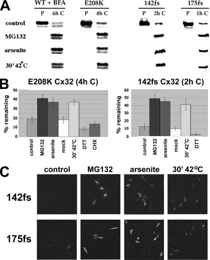
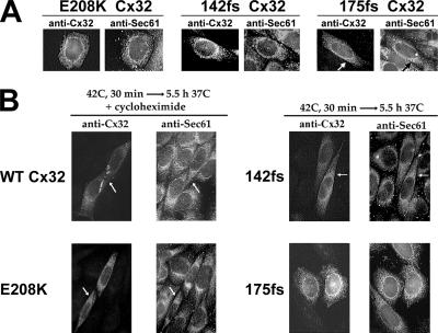
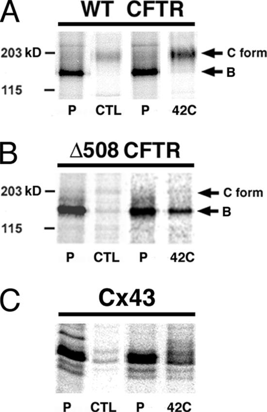
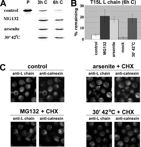
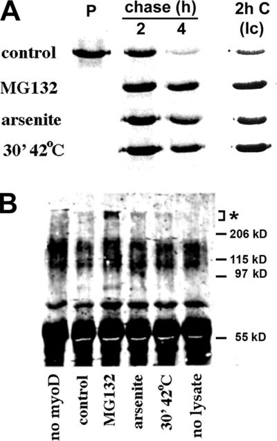
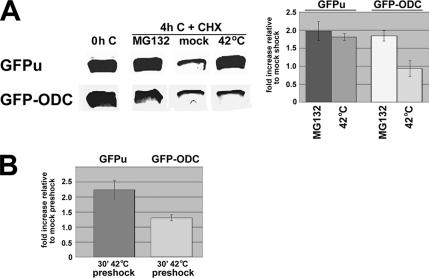
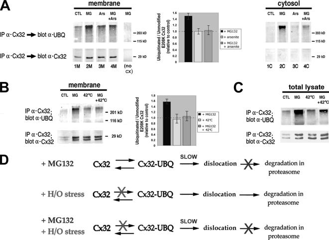
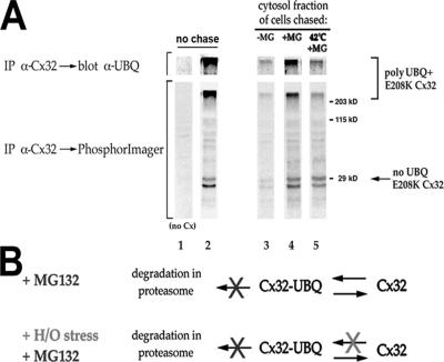
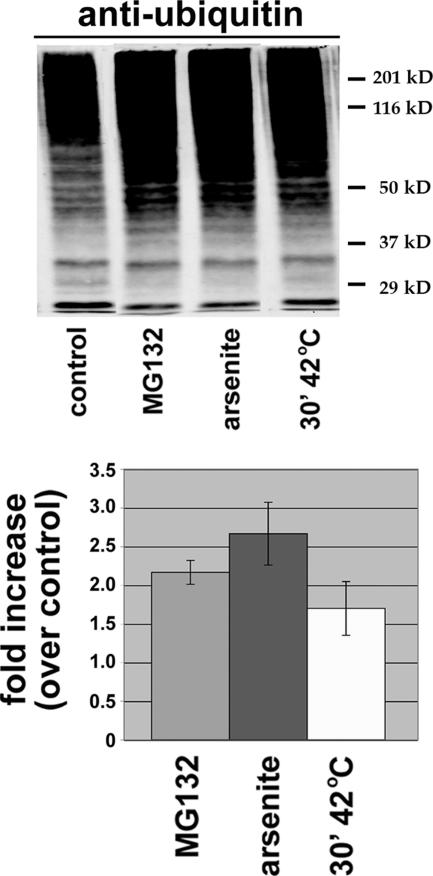
ER-associated, ubiquitin-proteasome system (UPS)-mediated degradation of the wild-type (WT) gap junction protein connexin32 (Cx32) is inhibited by mild forms of cytosolic stress at a step before its dislocation into the cytosol. We show that the same conditions (a 30-min, 42 degrees C heat shock or oxidative stress induced by arsenite) also reduce the endoplasmic reticulum (ER)-associated turnover of disease-causing mutants of Cx32 and the cystic fibrosis transmembrane conductance regulator (CFTR), as well as that of WT CFTR and unassembled Ig light chain. Stress-stabilized WT Cx32 and CFTR, but not the mutant/unassembled proteins examined, could traverse the secretory pathway. Heat shock also slowed the otherwise rapid UPS-mediated turnover of the cytosolic proteins myoD and GFPu, but not the degradation of an ubiquitination-independent construct (GFP-ODC) closely related to the latter. Analysis of mutant Cx32 from cells exposed to proteasome inhibitors and/or cytosolic stress indicated that stress reduces degradation at the level of substrate polyubiquitination. These findings reveal a new link between the cytosolic stress-induced heat shock response, ER-associated degradation, and polyubiquitination. Stress-denatured proteins may titer a limiting component of the ubiquitination machinery away from pre-existing UPS substrates, thereby sparing the latter from degradation.


Figures

Figure 1.
Figure 1.
Induction of H/O stress decreases ERAD of WT and mutant forms of Cx32. (A) CHO cells transiently transfected with plasmids encoding wild-type (WT), E208K, 142fs, or 175fs Cx32 were metabolically labeled with [35S]methionine at 37°C for 20 min and lysed either immediately (P) or after being chased for the indicated period with no additions (control), 30 μM MG132, 80 μM sodium arsenite, or for 30 min at 42°C immediately after the pulse and returned to 37°C for the remainder of the chase period. For WT Cx32 only, BFA was included in the pulse and chase media. Total cellular Cx32 was immunoprecipitated and analyzed by SDS-PAGE and PhosphorImaging. (B) The amount of [35S]methionine-Cx32 recovered by immunoprecipitation after a 4-h (E208K Cx32) or 2-h (142fs Cx32) chase under the indicated conditions was expressed as a percentage of [35S]methionine-Cx32 immunoprecipitable immediately after the pulse. Mock-shocked cells were treated identically to heat-shocked samples, except that the temperature was maintained at 37°C throughout the experiment. For each experimental condition, n = 3. (C) CHO cells transiently transfected to express 142fs or 175fs Cx32 were incubated for 4 h at 37°C with no additions (control), MG132, or arsenite as indicated. Heat-shocked cells were incubated at 42°C for 30 min and then returned to 37°C for an additional 3.5 h. All cells were then fixed and immunostained with anti-Cx32 antibodies.
Figure 2.
Figure 2.
H/O stress does not restore trafficking of mutant Cx32. CHO cells transiently transfected with plasmids encoding the indicated Cx32 species were incubated for 6 h at 37°C with no additions (A), or at 42°C for 30 min and then returned to 37°C for an additional 5.5-h incubation in either the presence (WT and E208K Cx32) or absence (175fs and 142fs Cx32) of cycloheximide (B). All cells were then fixed and doubly immunostained for Cx32 and the ER marker Sec61β.
Figure 3.
Figure 3.
H/O stress decreases the degradation of both WT and Δ508 CFTR, but only promotes the maturation of the former. CHO cells stably expressing either WT (A) or Δ508 (B and C) CFTR were either “preshocked” by a 30-min incubation at 42°C (42C), or mock-shocked at 37°C (CTL), and returned to 37°C for 4 h. The cells were then pulsed for 30 min with [35S]methionine and either lysed immediately (P) or chased for 6 h at 37°C (WT) or 27°C (Δ508) before immunoprecipitation of CFTR from the RIPA soluble cell lysate (A and B) or of endogenous Cx43 from the pooled RIPA soluble and insoluble fractions boiled in SDS (C). The contrast of the image of each pulse-chase pair was adjusted such that the intensity of all of the pulse (P) lanes was approximately equal.
Figure 4.
Figure 4.
H/O stress promotes the stability, but not the intracellular transport, of a soluble ERAD substrate. (A) SP2/0 myeloma cells stably expressing the unassembled, secretion-incompetent T15L immunoglobulin light chain were metabolically labeled for 20 min and lysed either immediately (P) or after being chased for the indicated period with no additions (control), MG132, sodium arsenite, or for 30 min at 42°C immediately after the pulse and returned to 37°C for the remainder of the chase period. The cells were treated with SDS, and immunoprecipitated with anti-L chain antibodies. (B) The amount of [35S]methionine-L chain recovered by immunoprecipitation from the cells after a 6-h chase under the indicated conditions was expressed as a percentage of [35S]methionine-L chain immunoprecipitable immediately after the pulse. For each experimental condition, n = 3 or 4. (C) The T15L-expressing cells were incubated for 4 h at 37°C with no additions (control) or for a total of 4 h in the presence of CHX + arsenite, CHX + MG132, or CHX for 30 min at 42°C followed by 3.5 h at 37°C. All cells were then fixed and immunostained with anti-L chain antibodies and antibodies specific for the ER marker calnexin.
Figure 5.
Figure 5.
Inhibition of degradation and polyubiquitination of the cytosolic/nuclear UPS substrate MyoD by H/O stress. (A) CHO cells transiently transfected with plasmids encoding MyoD were lysed and immunoprecipitated with anti-MyoD antibodies either immediately after a 20-min pulse with [35S]methionine (P) or after being chased for the indicated period with no additions (control), MG132, sodium arsenite, or for 30 min at 42°C immediately after the pulse and returned to 37°C for the remainder of the chase period. A lower contrast (lc) image of the 2-h chase data is included to indicate the extent to which MG132 and stress increase the recovery of MyoD at this time point; relative to the amount of [35S]methionine-MyoD recovered from control cells, the fold increase was: 2.09 ± 0.26 (MG132); 2.21 ± 0.62 (arsenite); and 3.65 ± 0.56 (hyperthermia). For each experimental condition, n = 3. (B) CHO cells transiently cotransfected with plasmids encoding MyoD and HA-tagged ubiquitin at a ratio of 1:5 were incubated for 4 h as in A before immunoprecipitation of MyoD. The samples were subjected to SDS-PAGE, transferred to PVDF membranes, and probed for polyubiquitinated MyoD (asterisk) with anti-HA antibodies. The no-MyoD sample was prepared from cells transfected with ubiquitin-HA only and treated as in lane 3; the no-lysate control shows the background signal from the immunoprecipitating antibody. Representative of three experiments. Anti-MyoD blots confirmed that the lack of polyubiquitinated MyoD from cells subjected to H/O stress was not due to decreased levels of MyoD protein.
Figure 6.
Figure 6.
Heat stress inhibits the degradation of EGFP bearing a ubiquitin-dependent degron, but not that of EGFP with a ubiquitin-independent degron. CHO cells were transiently transfected with plasmids encoding either GFPu or GFP-ODC. (A) Cells were lysed without any treatment (0h C), or incubated in the presence of cycloheximide for: 4 h at 37°C with MG132 (MG132), 30 min at 42°C followed by 3.5 h at 37°C (42°C), or 30 min at 37°C followed by 3.5 h at 37°C (mock). Total cell lysates were analyzed by Western blotting with anti-GFP antibodies. The amount of GFPu or GFP-ODC remaining after a 4-h chase in the presence of CHX under the indicated conditions was graphed as the fold increase relative to that obtained from CHX-treated mock-shocked cells. For each experimental condition, n = 3. (B) CHO cells transiently transfected with plasmids encoding either GFPu or GFP-ODC were “preshocked” by a 30-min incubation at 42°C or mock-shocked at 37°C and returned to 37°C for 3.5 h. The cells were then lysed without any treatment (0h C), or incubated for 4 h with CHX at 37°C and analyzed by Western blotting with anti-GFP antibodies. The amount of GFPu or GFP-ODC remaining after the CHX chase was calculated relative to the corresponding 0h C sample and was graphed relative to the value obtained from CHX-treated mock-shocked cells. For each experimental condition, n = 3.
Figure 7.
Figure 7.
Membrane-associated and total E208K Cx32 spared from ERAD by H/O stress is less highly ubiquitinated than E208K Cx32 saved by proteasome inhibitors. CHO cells were transiently cotransfected with plasmids encoding E208K Cx32 and HA-tagged ubiquitin at a ratio of 1:5. (A) The cells were incubated for 4 h without additions (control; CTL), or with MG132 (MG), arsenite (Ars), or both. All cells were then lysed without detergent and fractionated into cytosolic (C) and membrane (M) fractions before solubilization in SDS and immunoprecipitation of Cx32. Immunoprecipitates were subjected to SDS-PAGE on 12% gels and transferred to PVDF membranes. The blots were cut in half, and the lower portion was probed for unmodified E208K Cx32 with anti-Cx32 antibodies and the upper portion probed for polyubiquitinated E208K Cx32 with anti-ubiquitin antibodies. A “no cx” sample was prepared from cells transfected with ubiquitin-HA only and treated as in lane 2M. For each membrane sample from cells treated with MG132 and/or arsenite, the signal obtained for polyubiquitinated E208K Cx32 was calculated as a percentage of that obtained for polyubiquitinated E208K Cx32 from control cells in the same experiment. A similar calculation was performed for unmodified E208K Cx32 in the membrane. The ratio of the percentage obtained for polyubiquitinated E208K Cx32 to the percentage obtained for unmodified E208K Cx32 was calculated, and graphed relative to the ratio obtained from controls (defined as 100%/100%; 1). Data from three independent experiments. (B) Cotransfected cells were incubated in the presence of CHX for 2 h at 37°C with no other additions (control), 2 h at 37°C with MG132, 30 min at 42°C and returned to 37°C for 1.5 h, or 30 min at 42°C and returned to 37°C for 1.5 h in the continuous presence of MG132. Cells were lysed, and membrane fractions were analyzed as in A. Data from three independent experiments. (C) Cotransfected cells were incubated as in B, except that the CHX treatment was extended from 2 to 4 h. Cells were lysed in the presence of SDS, and whole cell lysates were analyzed for both polyubiquitinated and unmodified E208K Cx32. (D) Schematic of the effect of MG132 and/or H/O stress on ERAD of E208K Cx32. Top, proteasome inhibitors slow, but do not completely abolish, dislocation, and have a stronger inhibitory effect on proteolysis within the 20S proteasome core. Under these conditions, E208K Cx32 accumulates in the membrane in both ubiquitinated and nonubiquitinated forms and is also slowly dislocated into the cytosol after ubiquitination but is not degraded. Middle, cytosolic stress reduces ubiquitination of E208K Cx32, thereby inhibiting its dislocation and causing E208K Cx32 to accumulate in the membrane in a less ubiquitinated form than in the presence of proteasome inhibitors. Any E208K Cx32 that does becomes ubiquitinated and dislocated is degraded by the proteasome. Bottom, ubiquitination of membrane-associated E208K Cx32 is also reduced when cells are subjected to H/O stress in the presence of MG132. The latter blocks degradation of the small amount of E208K Cx32 that becomes dislocated, allowing it to be recovered from the medium.
Figure 8.
Figure 8.
H/O stress promotes the recovery of dislocated, MG132-stabilized E208K Cx32 in a nonubiquitinated form. (A) CHO cells transiently cotransfected with plasmids encoding E208K Cx32 and HA-tagged ubiquitin were metabolically labeled for 4 h in the presence of MG132. The cells were lysed without detergent, and a cytosolic fraction was prepared, either immediately after labeling (lane 2) or after chasing in the presence of CHX as follows: for 4 h with no other additions (lane 3), for 4 h with MG132 (lane 4), or for 30 min at 42°C followed by 3.5 h at 37°C in the continuous presence of MG132 (lane 5). Cx32 was immunoprecipitated from the cytosolic fraction, subjected to SDS-PAGE on 12% gels, and transferred to PVDF membranes. The membranes were analyzed for radiolabeled E208K Cx32 by phosphorImaging and were probed with anti-ubiquitin antibodies. Only the top portion of the anti-ubiquitin blot (Mr >200 kDa) showed specific anti-ubiquitin immunoreactivity and is included. Lane 1, sample prepared from cells transfected with ubiquitin-HA only and treated as in lane 2. (B) Schematic of the effect of H/O stress on post-dislocation pools of E208K Cx32. When degradation of dislocated, ubiquitinated E208K Cx32 (Cx32-UBQ) is blocked by MG132, E208K Cx32 accumulates in the cytosol in a less highly ubiquitinated form if the cells are also subjected to H/O stress due to inhibition of connexin reubiquitination by stress.
Figure 9.
Figure 9.
Total cellular ubiquitin conjugates are increased by H/O stress. CHO cells transiently cotransfected with plasmids encoding E208K Cx32 and HA-tagged ubiquitin were incubated for 4 h with no additions (control), MG132, sodium arsenite, or for 30 min at 42°C followed by 3.5 h at 37°C. Equal amounts of total, SDS-solubilized cell lysates were analyzed by Western blotting using anti-ubiquitin antibodies, and the results were quantitated as the fold increase in anti-ubiquitin immunoreactivity under the indicated conditions relative to untreated controls. For each experimental condition, n = 3.

Similar articles

Cited by

References

    1. Abel A., Bone L. J., Messing A., Scherer S. S., Fischbeck K. H. Studies in transgenic mice indicate a loss of connexin32 function in X-linked Charcot-Marie-Tooth disease. J. Neuropathol. Exp. Neurol. 1999;58:702–710. - PubMed
    1. Abu Hatoum O., Gross-Mesilaty S., Breitschopf K., Hoffman A., Gonen H., Ciechanover A., Bengal E. Degradation of myogenic transcription factor MyoD by the ubiquitin pathway in vivo and in vitro: regulation by specific DNA binding. Mol. Cell. Biol. 1998;18:5670–5677. - PMC - PubMed
    1. Bence N. F., Bennett E. J., Kopito R. R. Application and analysis of the GFPu family of ubiquitin-proteasome system reporters. Methods Enzymol. 2005;399:481–490. - PubMed
    1. Bond U., Agell N., Haas A. L., Redman K., Schlesinger M. J. Ubiquitin in stressed chicken embryo fibroblasts. J. Biol. Chem. 1988;263:2384–2388. - PubMed
    1. Breitschopf K., Bengal E., Ziv T., Admon A., Ciechanover A. A novel site for ubiquitination: the N-terminal residue, and not internal lysines of MyoD, is essential for conjugation and degradation of the protein. EMBO J. 1998;17:5964–5973. - PMC - PubMed

Publication types