. 2007 Nov;18(11):4222-31.
doi: 10.1091/mbc.e06-05-0416. Epub 2007 Aug 15.

FGF-2 release from the lens capsule by MMP-2 maintains lens epithelial cell viability

Affiliations

FGF-2 release from the lens capsule by MMP-2 maintains lens epithelial cell viability

Frederique M D Tholozan et al. Mol Biol Cell. 2007 Nov.

Abstract

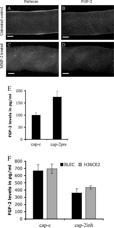
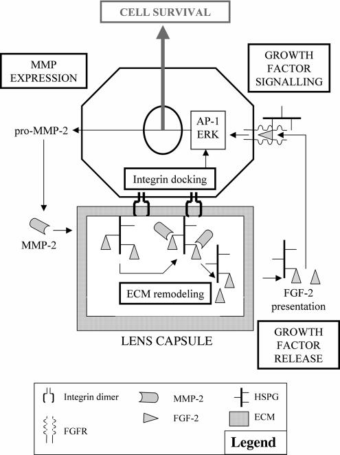
The lens is an avascular tissue, separated from the aqueous and vitreous humors by its own extracellular matrix, the lens capsule. Here we demonstrate that the lens capsule is a source of essential survival factors for lens epithelial cells. Primary and immortalized lens epithelial cells survive in low levels of serum and are resistant to staurosporine-induced apoptosis when they remain in contact with the lens capsule. Physical contact with the capsule is required for maximal resistance to stress. The lens capsule is also a source of soluble factors including fibroblast growth factor 2 (FGF-2) and perlecan, an extracellular matrix component that enhances FGF-2 activity. Matrix metalloproteinase 2 (MMP-2) inhibition as well as MMP-2 pretreatment of lens capsules greatly reduced the protective effect of the lens capsule, although this could be largely reversed by the addition of either conditioned medium or recombinant FGF-2. These data suggest that FGF-2 release from the lens capsule by MMP-2 is essential to lens epithelial cell viability and survival.


Figures

Figure 1.
Figure 1.
Lens epithelial cells are protected against serum deprivation when cultured on the lens capsule. BLEC and H36CE2 cells were seeded onto either plastic dishes (pl) or lens capsules (cap) in 0.1% (vol/vol) fetal cal serum (FCS). After 1 h in culture (A), there was no statistical difference (p > 0.05 for both BLEC and H36CE2) between the numbers of cells attached to the lens capsule or to the plastic dishes (bars show the average number of cells attached to the lens capsules as a percentage of the average number of cells attached to the plastic dishes). After 96 h in culture (B), viable cells were only detected on the lens capsules (cap). Those on plastic (pl) appeared not to have survived.
Figure 2.
Figure 2.
Lens epithelial cells are protected against staurosporine-induced apoptosis when cultured on the lens capsule. (A and B) BLEC cells (A) and H36CE2 cells (B) were grown on either uncoated plastic dishes (pl-c) or on bovine lens capsules (cap-c) in 10% (vol/vol) serum and then challenged with 0.5 μM staurosporine (pl-s; cap-s). After 48-h exposure to staurosporine, most of the BLEC (A, pl-s) and H36CE2 (B, pl-s) grown on the plastic dishes did not survive compared with controls (A, B; pl-c and pl-s; p < 0.05 for both BLEC and H36CE2). When grown on the lens capsules (A and B, cap-s), there was, however, a smaller decrease in cell number (A and B; cf. cap-c and cap-s; p > 0.05 and p < 0.05 for BLEC and H36CE2, respectively). (C–F) Immunofluorescence microscopy of BLEC cells labeled with both propidium iodide (red) and TUNEL staining (green) and grown on either glass coverslips (C and D) or on bovine lens capsules (E and F) in the absence (C and E) or presence (D and F) of 0.5 μM staurosporine in media containing 10% (vol/vol) serum. After 48 h in the presence of staurosporine, chromatin reorganization was frequently observed in the nuclei of cells grown on glass as detected by propidium iodide staining. These propidium iodide–positive nuclei were also largely TUNEL-positive, indicating that significant DNA damage has also occurred, as indicated by the yellow color of their nuclei (arrows). In contrast, cells grown on the bovine lens capsule were mostly not TUNEL positive and chromatin reorganization was also not usually seen (F). Scale bars, 10 μm. (G) BLEC cells grown on either glass coverslips (gl) or on bovine lens capsules (cap) in the absence (-c) or presence (-s) of 0.5 μM staurosporine in media containing 10% (vol/vol) serum were labeled with DAPI and then scored for the presence of apoptotic nuclei. The number of apoptotic cells after 48-h exposure to staurosporine was then expressed as percentage of total cell number as described in Materials and Methods. In the absence of staurosporine, the percentage of apoptotic cells was low for cells grown on both glass (gl-c) and lens capsule substrates (cap-c). With the addition of staurosporine, cells grown on the coverslips exhibited a very large increase in the number of apoptotic cells (gl-p and gl-s; p < 0.05). In contrast, cells grown on the lens capsule exhibited a much smaller increase in number of apoptotic cells (cap-s).
Figure 3.
Figure 3.
MMP-2 expression and activation are increased in H36CE2 cells grown on the lens capsule compared with H36CE2 cells grown on plastic. (A) Analysis of the TaqMan gene expression array data plotted as relative change (ΔΔCt) in transcript levels between extracts from an equal number of cells cultured for 48 h on the lens capsule (bars) and the plastic substrate (baseline). These data show that H36CE2 cells grown on the lens capsule consistently express more ADAMTS-4 and MMP-2 and less TIMP-1 and - 4 than H36CE2 grown on plastic. MMP-9 mRNA expression was not detected. (B and C) Conditioned medium from H36CE2 cells grown on the plastic (pl) and the lens capsule (cap) for 48 h were concentrated by centrifugation and analyzed by immunoblotting for the presence of MMP-2 (B) and MMP-9 (C). Human recombinant latent gelatinases (hrMMP-2 and -9; asterisks) were included as standards. MMP-2 was detected in the medium from cells grown on both substrates in serum-free medium. Serum-free medium was used to prevent BSA from obscuring the MMP-2 protein signal after sample concentration. In medium from the lens capsule cultures, MMP-2 was predominantly detected as the cleaved active form (upper arrowhead; B) with a further cleaved minor form (lower arrowhead), whereas in medium from plastic cultures MMP-2 was mainly detected in the latent form (asterisk). MMP-9 was not detected in medium from cells grown on either substrate in 0.1% (vol/vol) serum conditions.
Figure 4.
Figure 4.
Both attachment and soluble factors are involved in H36CE2 cells viability on the lens capsule. Controls, the maximum cell viability potential of H36CE2 cells grown directly on both plastic (pl) and the lens capsule (cap) was measured after 48 h in 0.1% (vol/vol) serum conditions. Coculture experiments, the effect of the lens capsule upon H36CE2 cell viability was measured in a coculture system in 0.1% (vol/vol) serum. In this coculture system, H36CE2 viability was measured for cells grown on an insert contained within a plastic dish well. This allowed for different combinations of lens capsule and H36CE2 cells to be added to the bottom chamber and for the effect of soluble factors upon the viability of the H36CE2 cells grown on the well insert to be determined. After 48 h in culture, in the absence of both cells and lens capsule in the bottom chamber, H36CE2 viability was very similar to that seen for the plastic control (cf. Coculture; ‘no cells in bottom chamber; no cap’ with Controls; pl; p > 0.05). Adding a lens capsule to the bottom chamber in the absence of any additional cells in this compartment was also unable to increase significantly the base level of cell viability for H36CE2 cells in the upper chamber (cf. Coculture, “no cells in bottom chamber, plus cap” with Controls, pl; p > 0.05). In contrast, adding both a lens capsule and H36CE2 cells to the bottom chamber significantly increased H36CE2 cell viability in the upper chamber (cf. Coculture, “with cells in bottom chamber; plus cap” with Controls; pl; p < 0.05). The addition of H36CE2 cells alone to the bottom chamber had no significant effect upon the viability of H36CE2 cells in the upper chamber (cf. Coculture, “with cells in bottom chamber; no cap” with Controls, pl; p > 0.05).
Figure 5.
Figure 5.
MMP-2 can release lens capsule-bound FGF-2 and perlecan and affect the amount of FGF-2 accessible to cells grown on the lens capsule. MMP-2 treatment of the lens capsule depletes capsule-bound perlecan and FGF-2 stores. (A–D) Bovine lens capsules cleaned of all primary cells were exposed to human recombinant MMP-2 for 72 h in substrate buffer (5 mM CaCl2, 50 mM Tris-HCl, pH 8.0) and then processed for immunofluorescence microscopy. In the untreated lens capsules, perlecan (green; A) and FGF-2 (red; B) were still concentrated at the outer and inner faces of the anterior lens capsule. In MMP-2–treated lens capsules, both layers were largely depleted [perlecan (green; C) and FGF-2 (red; D)]. Scale bars, 20 μm. (E) Bovine lens capsules cleaned of all primary cells were exposed to human recombinant MMP-2 for 72 h in substrate buffer (5 mM CaCl2, 50 mM Tris, pH 8.0), and levels of FGF-2 released into the medium were measured by ELISA. In the treated samples, levels of FGF-2 released into the medium were significantly increased with the addition of human recombinant MMP-2 (p < 0.05). (F) FGF-2 release from the lens capsule by H36CE2 and BLEC cells is reduced by MMP-2 inhibition. H36CE2 cells and BLEC cells were seeded onto lens capsules in 0.1% (vol/vol) FCS-supplemented medium and challenged with 100 mM OA-Hy as a selective inhibitor of MMP-2. After 48 h, MMP-2 (cap-c: cap-2inh) inhibition resulted in significant decreases in FGF-2 levels released into the medium as measured by ELISA (p < 0.05 for both BLEC and H36CE2).
Figure 6.
Figure 6.
MMP-2 treatment affects the surface architecture of the lens capsule. Bovine lens capsules cleaned of all primary cells were exposed to human recombinant MMP-2 for 72 h in substrate buffer (5 mM CaCl2, 50 mM Tris, pH 8.0) and prepared for SEM analysis as described in Materials and Methods. (A) In the untreated lens capsules, at low magnification the inner surface of the anterior capsule had a homogenous appearance with an intricate meshwork of surface material. At higher magnification (A, insert), this meshwork of fine filaments was seen to be locally arranged in a honeycomb pattern (A, inset, arrows). (B) In MMP-2–treated lens capsules, at low magnification the ECM surface had a similar general appearance but had lost the fine detail. The honeycomb surface structures were removed and condensed remnants appeared in their place (B, inset, arrows). Scale bars, 1 μm; Scale bars, (insets) 0.2 μm.
Figure 7.
Figure 7.
MMP-2 mediates lens epithelial cell viability via the release of soluble factors from the lens capsule. (A and B) BLEC (A) and H36CE2 (B) cells were grown in 0.1% (vol/vol) serum on lens capsules that had been exposed previously to human recombinant MMP-2 for 72 h (cap-2pre). After 48 h in culture, this pretreatment caused a significant decrease in cell viability (A and B: cap-c and cap-pre; p < 0.05 for both BLEC and H36CE2). This decrease was not significantly different from the decrease in cell viability obtained when either BLEC or H36CE2 cells were exposed to the MMP-2 selective inhibitor OA-Hy for the 48-h culture time (A and B: cap-c and cap-2inh; p < 0.05 for both BLEC and H36CE2 and A and B: cap-2pre and cap-2inh; p > 0.05 for both BLEC and H36CE2). The addition of conditioned medium from MMP-2 pretreated lens capsules (CM) to either BLEC or H36CE2 cells grown on lens capsules in the presence of MMP-2 selective inhibitor OA-Hy (A and B: cap-2inh+CM) partially reversed the decrease in lens epithelial cell viability induced by the addition of selective MMP-2 inhibitor (A and B: cap-2inh and cap-2inh+CM; p < 0.05 for both BLEC and H36CE2, and A and B: cap-c and cap-2inh+CM; p > 0.05 and p < 0.05 for BLEC and H36CE2, respectively). (C) Exposure to MMP inhibitor II as a selective MMP-9 inhibitor failed to decrease the cell viability of either BLEC or H36CE2 cells grown on lens capsules in 0.1% (vol/vol) serum (cap-c and cap-9inh; p > 0.05 for both BLEC and H36CE2). (D) BLEC grown on the lens capsules were exposed to 100 μM MMP2-selective inhibitor OA-Hy (cap-2inh) in 0.1% (vol/vol) serum-supplemented medium. The subsequent decrease in cell viability (cap-c and cap-2inh) could be significantly, but not completely, reversed by the single addition of recombinant FGF-2 (200 pg/ml; cap-2inh and cap-2inh+FGF, p < 0.05, and cap-c and cap-2inh+FGF, p < 0.05).
Figure 8.
Figure 8.
The MMP-ECM-Growth factor (MEG) cycle for cell viability. The presence and activity of MMP-2 in the lens cells' environment, likely after integrin-mediated gene transcription as described for other systems (Troussard et al., 1999; Lee et al., 2005), leads to the degradation of HSPGs in the ECM, allowing soluble FGF-2 release into the media. FGF-2 then binds its receptor and activates downstream signaling pathways (Lovicu and McAvoy, 2001; Chandrasekher and Sailaja, 2003; Iyengar et al., 2007) to promote cell survival. This represents the simplest form of the cycle and does not exclude contributions from other proteinases, cryptogenic ECM sites, and IGF-1 and TGF-β2 release that can all influence epithelial cell survival. In other systems, FGF-2 can feedback to increase gelatinase expression (El Ramy et al., 2005; Wang et al., 2005).

Similar articles

Cited by

References

    1. Adams J. C., Watt F. M. Regulation of development and differentiation by the extracellular matrix. Development. 1993;117:1183–1198. - PubMed
    1. Andley U. P., Rhim J. S., Chylack L. T., Jr, Fleming T. P. Propagation and immortalization of human lens epithelial cells in culture. Invest. Ophthalmol. Vis. Sci. 1994;35:3094–3102. - PubMed
    1. Azuma N., Hara T. Extracellular matrix of opacified anterior capsule after endocapsular cataract surgery. Graefes Arch. Clin. Exp. Ophthalmol. 1998;236:531–536. - PubMed
    1. Bergers G., Brekken R., McMahon G., Vu T. H., Itoh T., Tamaki K., Tanzawa K., Thorpe P., Itohara S., Werb Z., Hanahan D. Matrix metalloproteinase-9 triggers the angiogenic switch during carcinogenesis. Nat. Cell Biol. 2000;2:737–744. - PMC - PubMed
    1. Bissell M. J., Radisky D. C., Rizki A., Weaver V. M., Petersen O. W. The organizing principle: microenvironmental influences in the normal and malignant breast. Differentiation. 2002;70:537–546. - PMC - PubMed

MeSH terms

LinkOut - more resources