. 2007 Oct;18(10):4155-67.
doi: 10.1091/mbc.e07-02-0094. Epub 2007 Aug 15.

Regulation of the formin for3p by cdc42p and bud6p

Affiliations

Regulation of the formin for3p by cdc42p and bud6p

Sophie G Martin et al. Mol Biol Cell. 2007 Oct.

Abstract

Formins are conserved actin nucleators responsible for the assembly of diverse actin structures. Many formins are controlled through an autoinhibitory mechanism involving the interaction of a C-terminal DAD sequence with an N-terminal DID sequence. Here, we show that the fission yeast formin for3p, which mediates actin cable assembly and polarized cell growth, is regulated by a similar autoinhibitory mechanism in vivo. Multiple sites govern for3p localization to cell tips. The localization and activity of for3p are inhibited by an intramolecular interaction of divergent DAD and DID-like sequences. A for3p DAD mutant expressed at endogenous levels produces more robust actin cables, which appear to have normal organization and dynamics. We identify cdc42p as the primary Rho GTPase involved in actin cable assembly and for3p regulation. Both cdc42p, which binds at the N terminus of for3p, and bud6p, which binds near the C-terminal DAD-like sequence, are needed for for3p localization and full activity, but a mutation in the for3p DAD restores for3p localization and other phenotypes of cdc42 and bud6 mutants. In particular, the for3p DAD mutation suppresses the bipolar growth (NETO) defect of bud6Delta cells. These findings suggest that cdc42p and bud6p activate for3p by relieving autoinhibition.


Figures
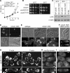
Figure 1.
Figure 1.
Cdc42p is necessary for cell morphology, actin cable organization and for3p localization. (A) Growth curve of wild-type and cdc42-1625 cells incubated at 28 and 36°C. (B) Rescue of the temperature-sensitive growth defect of cdc42-1625 by different cdc42 alleles. cdc42-1625 cells were transformed with the expression plasmid pREP41-HA carrying either no insert, wild-type cdc42+, constitutively active cdc42G12V, or dominant-negative cdc42T17N alleles and grown on Edinburgh minimal media plates for 2 d at 25 and 36°C. (C) Immunoblot of HA-cdc42p and HA-cdc42p-1625 expressed from the endogenous promoter. Twenty-five micrograms of total yeast extracts from exponential cultures at 25 and 36°C were loaded in each lane. Levels were monitored with the anti-actin antibody. Although wild-type cdc42p shows increased levels at 36°C, this increase failed to happen in the cdc42-1625 mutant. (D) Immunofluorescence of HA-cdc42p and HA-cdc42p-1625 with anti-HA antibody. Both wild-type and mutant cdc42p localize to growing cell tips. Differential interference contrast (DIC) of the permeabilized cells is shown at the bottom. (E) Morphology of wild-type and cdc42-1625 cells grown to log phase at 28 and 36°C for 5 h. DIC and calcofluor-staining images are shown. (F) Rescue of cell morphology (DIC; top) and actin cables (AlexaFluor-phalloidin; bottom) by wild-type cdc42+ in cdc42-1625 cells at 36°C. (G) Projection images of spinning disk confocal stacks of AlexaFluor 488-phalloidin–stained cdc42-1625 (top) and wild-type (bottom) cells grown at 25°C (left) or 36°C for 1 h (right). Note that actin cables are extremely weak, but they were still present in cdc42-1625 cells (arrowheads). (H) Single focal plane widefield fluorescence images of myo52p-tomato in cdc42-1625 (top) and wild-type (bottom) cells grown at 25°C. (I) Single focal plane widefield fluorescence images of for3p-3GFP in cdc42-1625 (top) and wild-type (bottom) cells grown at 25°C. Note that for3p largely fails to localize to cell tips in the mutant cells. All bars, 2 μm.
Figure 2.
Figure 2.
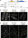
For3p contains three independent localization domains. (A) Scheme of for3p fragments that were fused to GFP to assay localization. All constructs were expressed from a plasmid under control of the medial-strength nmt1 promoter. (B) Projection images of spinning disk confocal stacks of GFP-for3N (left), GFP-for3(137-515) (middle), and GFP-for3NΔ(353-406) (right). Yellow arrowheads indicate cell tips and septum localization. GFP-for3N is also detected at the spindle pole body. (C) Projection images of spinning disk confocal stacks of GFP-for3C (left), GFP-for3C treated with 200 μM LatA (middle), and GFP-for3C-I930A (right). Blue arrowhead shows actin cable localization. Yellow arrowheads indicate cell tips and septum localization.
Figure 3.
Figure 3.
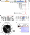
Identification of a DAD-like region in for3p necessary for interaction with the N terminus. (A) Mapping of the DAD domain by two-hybrid assay. Interaction of C-terminal for3p fragments cloned in the pGBD vector with pGAD-for3N(1-702) was assayed by growth on −His plates. (B) Sequence alignment of for3p DAD with defined DAD domains from S. cerevisiae, Drosophila, and mouse formins. Stretches of basic residues are shown in blue. Mutation of the residues indicated in red to alanines abolished interaction with for3p N terminus. (C) Two-hybrid assay on SC-His plate of pGAD-for3N(1-702), pGAD-bud6C(581-1385) and empty pGAD with wild-type, LLT-AAA, and RKK-AAA pGBD-for3C(1261-1461). Numbers refer to constructs indicated in A. Mutations in the DAD region specifically abolish interaction with for3N but not with bud6C. (D) 6His-for3C binds directly and specifically to MBP-for3N. Binding of bacterially expressed proteins was assayed by affinity column. 6His-for3C (aa 630-1461) binds to MBP-for3N(1-702), but not MBP alone. This interaction is compromised by the LLT-AAA mutation (DAD*). 6His-tagged protein fragments were detected by Western blotting with an anti-6His antibody. Coomassie staining of MBP-fusions indicates equal loading.
Figure 4.
Figure 4.
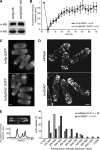
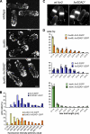
Phenotype of the for3DAD* mutant. (A) Immnunoblot of for3p-HA and for3pDAD*-HA expressed from the endogenous promoter. Twelve micrograms of total yeast extract were loaded in each lane. Levels were monitored with the TAT anti-α-tubulin antibody. (B) FRAP analysis of for3p-3GFP and for3pDAD*-2GFP. Using a laser scanning microscope, the entire cell tip of cells expressing either for3p-3GFP or for3pDAD*-2GFP from the endogenous promoter was photobleached and imaged every 4 s thereafter to monitor fluorescence recovery. Each trace represents the average value for the indicated number of experiments. Error bars represent the SE. (C) Single focal plane widefield fluorescence images of for3p-3GFP and for3pDAD*-2GFP expressed from the endogenous promoter. Note that for3pDAD* localizes to cell tips, if not even more tightly than wild type for3p. (D) Projection images of spinning disk confocal stacks of AlexaFluor 488-phalloidin stained wild-type (top) and for3DAD* (bottom) cells. Note that actin cables in the for3DAD* mutant stain more brightly than in wild-type cells. (E) Quantification of the fluorescence intensity of actin cables in wild-type and for3DAD* cells. We measured the peaks of the fluorescence profile along a line drawn across actin cables and subtracted background value. A histogram of these values is shown. Note that the fluorescence value is arbitrary and varies from one experiment to the other, but that under identical staining and imaging conditions, for3DAD* cells always show stronger actin cable staining than wild-type cells.
Figure 5.
Figure 5.
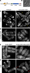
Deletion within the for3p DID leads to disorganized actin cables. (A) Scheme of the Δ353-406 deletion within full-length for3p. (B) DIC image of for3Δ353-406-myc cells. (C) Projection images of spinning disk confocal stacks of AlexaFluor 488-phalloidin–stained wild-type and for3Δ(353-406)-myc cells. Note the disorganized appearance of actin cables in for3Δ(353-406)-myc cells. Yellow arrowheads indicate locations from which some actin cables seem to radiate. (D) Single focal plane widefield fluorescence images of myo52p-GFP in wild-type (left) and for3Δ(353-406)-myc (right) cells. (E) Single focal plane widefield fluorescence images of for3p-3GFP and for3pΔ(353-406)-2GFP (right). (F) Single focal plane widefield fluorescence images of for3p-3GFP (left) and for3pΔ(353-406)-2GFP (right) in cells treated with 200 μM LatA. Red arrowheads highlight cell tip localization of for3pΔ(353-406)-2GFP.
Figure 6.
Figure 6.
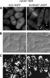
Cdc42p relieves for3p autoinhibition to regulate its localization. (A) Single focal plane widefield fluorescent image of for3p-3GFP (left) and for3pDAD*-2GFP (right) in cdc42-1625 cells. Note that the DAD* mutation is sufficient to restore for3p localization to cell tips in cdc42-1625 mutant cells (arrowheads). (B) DIC images of for3-3GFP cdc42-1625 (left) and for3DAD*-2GFP cdc42-1625 (right) cells. The DAD* mutation improves the morphology of cdc42-1625 mutant cells. (C) Projection images of spinning disk confocal stacks of AlexaFluor 488-phalloidin–stained for3-3GFP cdc42-1625 (left) and for3DAD*-2GFP cdc42-1625 (right) cells. Arrowheads point at weak actin cables. Note that the DAD* mutation fails to restore wild-type actin cables in cdc42-1625 mutant cells.
Figure 7.
Figure 7.
Bud6p targets for3p to cell tips through both anchoring and relief of autoinhibition. (A) Mapping of the BBS in for3p by two-hybrid analysis. Interaction of C-terminal for3p fragments cloned in the pGBD vector with pGAD-bud6C(581-1385) was assayed by growth on −His plates. Note how the minimum interaction domain overlaps with the DAD region. (B) Single focal plane widefield fluorescent images of for3p-3GFP in bud6Δ cells. Cells on the right were treated with 200 μM LatA. (C) Single focal plane widefield fluorescent images of for3pDAD*-2GFP bud6Δ cells. Cells on the right were treated with 200 μM LatA. Note that the DAD* mutation is sufficient to restore efficient for3p localization to cell tips in bud6Δ mutant cells (blue arrowheads). (D) Projection images of spinning disk confocal stacks of GFP-for3N (left) and GFP-for3C (middle), and GFP-for3C-I930A (right) expressed from the medial-strength nmt1 promoter in bud6Δ mutant cells. GFP-for3C fails to localize to cell tips. (E) Single focal plane widefield fluorescent images of cdc42p-GFP in wild-type and bud6Δ cells. (F) Single focal plane widefield fluorescent images of bud6p-GFP in wild-type and cdc42-1625 cells.
Figure 8.
Figure 8.
Mutation of the for3p DAD domain restores wild-type actin cables and bipolar growth in bud6Δ mutants. (A) Projection images of spinning disk confocal stacks of AlexaFluor 488-phalloidin–stained for3-3GFP (wild-type), bud6Δ for3-3GFP (bud6Δ), and bud6Δ for3DAD*-2GFP (bud6Δ for3DAD*) cells. Note that the intensity of actin cables is lower in bud6Δ cells compared with wild-type cells, but that this phenotype is suppressed by the DAD* mutation. (B) Quantification of the fluorescence intensity of actin cables in for3-3GFP, for3DAD*-2GFP, bud6Δ for3-3GFP, and bud6Δ for3DAD*-2GFP cells, as in Figure 4E. (C) Calcofluor staining of bud6Δ cells expressing either for3-3GFP (wt for3) or for3DAD*-2GFP (for3DAD*), showing monopolar and bipolar growth, respectively. (D) Quantification of new end growth in strains of the indicated phenotypes. The length between the inner side of the birth scar (black line in the calcofluor staining) and the tip of the cell was measured, as shown in C. This length is of up to 1.5 μm in cells that fail to initiate growth at the new end.

Similar articles

Cited by

References

    1. Alberts A. S. Identification of a carboxyl-terminal diaphanous-related formin homology protein autoregulatory domain. J. Biol. Chem. 2001;276:2824–2830. - PubMed
    1. Cadwell R. C., Joyce G. F. Randomization of genes by PCR mutagenesis. PCR Methods Appl. 1992;2:28–33. - PubMed
    1. Chang F., Drubin D., Nurse P. cdc12p, a protein required for cytokinesis in fission yeast, is a component of the cell division ring and interacts with profilin. J. Cell Biol. 1997;137:169–182. - PMC - PubMed
    1. Dong Y., Pruyne D., Bretscher A. Formin-dependent actin assembly is regulated by distinct modes of Rho signaling in yeast. J. Cell Biol. 2003;161:1081–1092. - PMC - PubMed
    1. Eisenmann K. M., Harris E. S., Kitchen S. M., Holman H. A., Higgs H. N., Alberts A. S. Dia-interacting protein modulates formin-mediated actin assembly at the cell cortex. Curr. Biol. 2007;17:579–591. - PubMed

Publication types

MeSH terms