. 2007 Oct;18(10):4129-42.
doi: 10.1091/mbc.e07-01-0080. Epub 2007 Aug 15.

Rab6 regulates both ZW10/RINT-1 and conserved oligomeric Golgi complex-dependent Golgi trafficking and homeostasis

Affiliations

Rab6 regulates both ZW10/RINT-1 and conserved oligomeric Golgi complex-dependent Golgi trafficking and homeostasis

Yi Sun et al. Mol Biol Cell. 2007 Oct.

Abstract

We used multiple approaches to investigate the role of Rab6 relative to Zeste White 10 (ZW10), a mitotic checkpoint protein implicated in Golgi/endoplasmic reticulum (ER) trafficking/transport, and conserved oligomeric Golgi (COG) complex, a putative tether in retrograde, intra-Golgi trafficking. ZW10 depletion resulted in a central, disconnected cluster of Golgi elements and inhibition of ERGIC53 and Golgi enzyme recycling to ER. Small interfering RNA (siRNA) against RINT-1, a protein linker between ZW10 and the ER soluble N-ethylmaleimide-sensitive factor attachment protein receptor, syntaxin 18, produced similar Golgi disruption. COG3 depletion fragmented the Golgi and produced vesicles; vesicle formation was unaffected by codepletion of ZW10 along with COG, suggesting ZW10 and COG act separately. Rab6 depletion did not significantly affect Golgi ribbon organization. Epistatic depletion of Rab6 inhibited the Golgi-disruptive effects of ZW10/RINT-1 siRNA or COG inactivation by siRNA or antibodies. Dominant-negative expression of guanosine diphosphate-Rab6 suppressed ZW10 knockdown induced-Golgi disruption. No cross-talk was observed between Rab6 and endosomal Rab5, and Rab6 depletion failed to suppress p115 (anterograde tether) knockdown-induced Golgi disruption. Dominant-negative expression of a C-terminal fragment of Bicaudal D, a linker between Rab6 and dynactin/dynein, suppressed ZW10, but not COG, knockdown-induced Golgi disruption. We conclude that Rab6 regulates distinct Golgi trafficking pathways involving two separate protein complexes: ZW10/RINT-1 and COG.


Figures
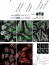
Figure 1.
Figure 1.
Treatment of HeLa cells with siRNAs directed against ZW10 or Rab6 effectively depleted each with little effect on the processing of LAMP2, even though the organization of the Golgi ribbon was affected. HeLa cells stably expressing GalNAcT2-GFP were transfected with either scrambled siRNA (control), siZW10(102), or siRab6(554) at a concentration of 200 nM in the absence of fetal bovine serum for 4 h and then cultured for 72 h. (A) Western blotting using affinity-purified antibodies to human ZW10 showed extensive knockdown of ZW10 relative to GAPDH as control. Similarly, Rab6 was extensively depleted. Under these conditions, there was no detectable decrease in the extensive Golgi glycosylation of LAMP2, and, in Rab6 siRNA treatment, a small increase in glycosylation as indicated by decreased mobility. (B–D) Fluorescence characterization of the distribution of GalNAcT2-GFP (green) indicated that siZW10 and siRab6 treatment had contrasting effects on the organization of the Golgi ribbon with little, if any, effect on the arrangement of microtubules (MT; white) or general cell shape. Asterisk in C, siZW10(102) marks an example of the occasional nondisrupted Golgi apparatus (∼5%) seen in cells treated with siRNA directed against ZW10. (E and F) Normal distribution of ER exit sites (Sec13a; white) in HeLa cells treated with siScrambled (E) or siZW10(102) (F) siRNAs for 72 h and stained for endogenous GalT (red). Images shown in B–F are all maximum intensity projections of confocal image stacks through the full cell depth. These images were taken with a 63×/1.40 numerical aperture objective. (G and H) Photobleaching and quantification was done as described under Materials and Methods at room temperature. Arrowheads in G point to ∼1-μm2 areas bleached and quantified as illustrated in H for several examples.
Figure 2.
Figure 2.
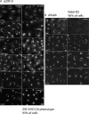
Effect of siRNAs directed against ZW10 and Rab6 on the distribution of a wide range of Golgi proteins. (A and B) Wild-type HeLa cells were siRNA treated for 72 h as described in the legend to Figure 1, and then they were stained for various endogenous Golgi-associated proteins (Kinesin, Rab6, GRASP55, GRASP65, p115, and GM130. GalT). Micrographs taken with a 63×/1.4 numerical aperture objective under identical exposure conditions for both scrambled siRNA-treated cells and cells treated with either siZW10(102) or Rab6(554). All micrographs shown in A and B are single plane projections of full-cell, confocal image stacks.
Figure 3.
Figure 3.
siRNAs directed against ZW10 had little, if any, effect on minus- and plus-end–directed movement of Golgi elements or late endosomes/lysosomes; in contrast, siRNAs directed against Rab6 induced plus-end accumulation of late endosomes/lysosomes at the cell periphery. siRNA treatments were for 72 h as described in the legend to Figure 1. All images shown are full-cell confocal image projections into a single plane. (A) A′ and B′, transport (minus-end–directed transport) of tsO45G-GFP protein from ER to clustered Golgi elements in siZW10(102) occurred in the normal 30-min time frame. Cells are stained for GalT (red). (B) A′ and B′, siZW10(201) treatment for 72 h had had minor effects on the distribution of LAMP2, a late endosomal/lysosome marker. (C) Treatment of GalNAcT2-GFP HeLa cells were siRab6(554) dispersed LAMP2 to toward the cell periphery. Arrows point to LAMP2 concentrations in cell tips. (D) A′–I′, the Golgi apparatus in both siZW10(102)- and siRab6(554)-treated cells is sensitive to BFA (plus-end–directed transport) and Golgi reassembly occurred after BFA washout and transport (minus-end–directed transport) of GalNAcT2-GFP from the ER.
Figure 4.
Figure 4.
siRNAs directed against ZW10 inhibited the cycling of ERGIC53 and Golgi enzymes to the ER. (A) Effect of 200 nM siZW10(102) treatment for 72 h on the distribution of ERGIC53. siScrambled (A′) and siZW10(102) (B′), 72 h. Full-cell confocal image projections into a single plan. (B) Effect of ZW10 knockdown on the constitutive recycling of a Golgi enzyme, GalNAcT2, to the ER as revealed by a Sar1p ER exit block. HeLa cells stably expressing GalNAcT2-GFP were treated with 200 nM ZW10(102) siRNA for 70 h, two cycles of transfection. Control and siZW10 cells were microinjected with a pCMUIV plasmid encoding GDP-restricted Sar1p. Five hours later, cells were fixed, and the distribution of GalNAcT2 was scored on an individual cell basis. In absence of any block, ER-like distribution of GalNAcT2 was rare (no block). In the siZW10 cells, there was a 3 times inhibition. Asterisks in A′ (control) and B′ (siZW10) mark examples of injected cells. Widefield micrographs.
Figure 5.
Figure 5.
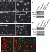
Rab6 depletion, GDP-Rab6a overexpression, or overexpression of BicD C-fragment, a Rab6 effector blocker, inhibit ZW10(102) siRNA-induced dispersal of the Golgi apparatus. (A–D) Cells double-treated with Rab6 + ZW10(102) siRNAs show compact Golgi phenotype. HeLa cells stably expressing GalNAcT2-GFP were mock (scrambled siRNA) (A) or ZW10(102) (B), Rab6 (C), and double (Rab6 + ZW10) (D) siRNA treated for 72 h and then fixed. Widefield images. (E) Total cell lysates from A to D were immunoblotted with anti-ZW10 (top row), anti-Rab 6 (middle row), and anti-tubulin (bottom row) antibodies. (F–H) Rab5 siRNA knockdown does not interfere with ZW10(102) siRNA-induced unraveling of Golgi. HeLa cells stably expressing GalNAcT2-GFP were Rab5 (F) or double (Rab5 + ZW10) (G) siRNA treated. (H) Total cell lysates from F and G were immunoblotted with anti-ZW10, anti-Rab5, and anti-tubulin as a loading control. (I) A′, GDP-Rab6a overexpression inhibited display of the siZW10 Golgi phenotype. GalNAcT2-GFP HeLa cells were transfected with ZW10 siRNA for 57 h, and then plasmids encoding GDP-Rab6 (100 ng/μl) were microinjected into cell nuclei. Six hours later, cells were fixed and stained with anti-Rab6 antibody (red). In the injected cells (red), the Golgi apparatus has a compact, juxtanuclear distribution. Strong inhibition of the ZW10 knockdown phenotype was seen in 60% of the microinjected cells. Widefield images. B′ and C′, expression of BicD C-fragment inhibits Golgi dispersal in response to ZW10 depletion. B′, control GalNAcT2-GFP HeLa cells were microinjected with plasmids (80 ng of DNA/μl stock concentration) encoding BicD C-fragment (myc tagged), and after a 24-h expression period they were myc stained (red). Expression of BicD C-fragment had no effect on the organization of the Golgi ribbon. C′, HeLa cells stably expressing GalNAcT2-GFP were incubated for 48 h after transfection with ZW10(102) siRNA. At 24 h post-siRNA transfection, cells were microinjected with plasmids (60–200 ng DNA/μl encoding BicD C-fragment; myc tagged). At the end of the 24-h expression period, cells were fixed and stained with anti-myc antibody to identify BicD-C–positive cells (red). BicD-C cells, 50% incidence, frequently showed a compact, juxtanuclear Golgi apparatus in comparison with the unraveled Golgi apparatus normally seen with ZW10 knockdown. Widefield images.
Figure 6.
Figure 6.
Rab6 depletion inhibits RINT-1 siRNA-induced dispersal of the Golgi apparatus. (A–D) Cell double-treated with Rab6 + RINT-1 siRNAs shows compact Golgi phenotype. HeLa cells stably expressing GalNAcT2-GFP were control (scrambled siRNA) (A) or RINT-1 (B), Rab6 (C), and double (Rab6 + RINT-1) (D) siRNA treated for 72 h and then fixed. Micrographs are maximum intensity projection of confocal image stacks. (E) Total cell lysates were immunoblotted with anti-RINT-1 (top row), anti-Rab6 (middle row), and anti-tubulin (bottom row) antibodies.
Figure 7.
Figure 7.
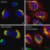
HeLa cells double-treated with COG3 and ZW10 siRNAs display a phenotype that shows vesicular accumulation of GlcNAcT1 as observed with COG3 siRNA treatment. HeLa cells stably expressing GlcNAcT1-myc (red channel) were mock (control, A) or COG3 (B). ZW10 (C) and double (ZW10 + COG3) (D) siRNA treated for 72 h, 100 nM individual duplex siRNA concentration, fixed, and stained with anti-myc antibody, anti-GM130 (green channel); and DNA (DAPI, blue channel). Image stacks were taken with the CARV II spinning disk confocal accessory and projected into a single XY plane.
Figure 8.
Figure 8.
Rab6 depletion inhibits COG3 siRNA-induced fragmentation of Golgi and accumulation of GlcNAcT1 in vesicles. (A–D) Cells double-treated with Rab6 + COG3 siRNAs show compact Golgi phenotype. HeLa cells stably expressing GlcNAcT1-myc were mock (control) (A) or COG3 (B), Rab6 (C), and double (Rab6 + COG3) (D) siRNA treated for 72 h, fixed, and stained with anti-myc antibody. Myc signal was collected as confocal image stacks by using a CARV II confocal accessory. (E) Total cell lysates from A to D were immunoblotted with anti-Cog3 (top row), anti-Rab 6 (middle row), and anti-GAPDH (bottom row) antibodies. (F–H) Rab5 siRNA knockdown does not interfere with COG3 siRNA-induced fragmentation of Golgi. HeLa cells stably expressing GalNAcT2-GFP were COG3 (F) or double (Rab5/COG3) (G) siRNA treated. (H) Total cell lysates from F and G were immunoblotted with anti-Rab5 and anti-β-actin as a loading control. Efficiency of knockdown was ∼90%. Widefield images. Arrows indicate fragmented Golgi apparatus. Bar, 10 μm.
Figure 9.
Figure 9.
Rab6 knockdown reduces Golgi fragmentation caused by microinjection of Cog3 antibody. HeLa cells stably expressing GalNAcT2-GFP were mock (A and B) or Rab6 (C and D) siRNA treated for 72 h. Cells were microinjected with affinity-purified anti-Cog3 (A and C) or preimmune (B and D) antibodies and fixed 4 h after microinjection. GFP signal was collected as confocal image stacks by using a CARV II confocal accessory. Arrow, fragmented Golgi; arrowhead, compact Golgi; asterisk, injected cells. Bar, 10 μm.
Figure 10.
Figure 10.
Rab6 knockdown fails to inhibit Golgi fragmentation in response to p115 depletion. HeLa cells stably expressing GalNAcT2-GFP were treated for 72 h with control scrambled siRNA (A), p115 siRNA (B), Rab6 siRNA (C), or p115 + Rab6 siRNAs (D). Widefield images. (E) Immunoblot showing that p115 and Rab6 are appropriately knocked down.
Figure 11.
Figure 11.
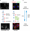
Schematic model of the initiating role of Rab6 in ZW10/RINT-1– and COG-dependent Golgi trafficking pathways necessary for normal Golgi homeostasis. Our data suggest that a role of Rab6 is in the initiation of two Golgi recycling pathways: recycling of Golgi components to the ER in pathway requiring a BicD-dependent motor and ZW10/RINT-1 (A) and intra-Golgi recycling of Golgi enzymes in a COG-dependent pathway (B). Rab6 has no effect on p115 anterograde trafficking. Both pathways are required for normal Golgi homeostasis. (C) Balance of the various proteins relative to Golgi adherence versus dispersal.

Similar articles

Cited by

References

    1. Allan B. B., Moyer B. D., Balch W. E. Rab1 recruitment of p115 into a cis-SNARE complex: programming budding COPII vesicles for fusion. Science. 2000;289:444–448. - PubMed
    1. Andag U., Schmitt H. H. Dsl1p, an essential component of the Golgi-endoplasmic reticulum retrieval system in yeast, uses the same sequence motif to interact with different subunits of the COPI vesicle coat. J. Biol. Chem. 2003;278:51722–51734. - PubMed
    1. Antony C., Cibert C., Geraud G., Santa Maria A., Maro B., Mayau V., Goud B. The small GTP-binding protein rab6p is distributed from medial Golgi to the traan-Golgi network as determined by a confocal microscopic approach. J. Cell Sci. 1992;103:785–796. - PubMed
    1. Arasaki K., Taniguchi M., Tani K., Tagaya M. RINT-1 regulates the localization and entry of ZW10 to the syntaxin 18 complex. Mol. Biol. Cell. 2006;17:2780–2788. - PMC - PubMed
    1. Burkhardt J. K., Echeverri C. J., Nilsson T., Vallee R. B. Overexpression of the dynamitin (p50) subunit of the dynactin complex disrupts dyein-dependent maintenance of membrane organelle distribution. J. Cell Biol. 1997;139:469–484. - PMC - PubMed

Publication types

MeSH terms

LinkOut - more resources