. 2007 Oct;18(10):4096-105.
doi: 10.1091/mbc.e06-11-0993. Epub 2007 Aug 15.

Multiple myosins are required to coordinate actin assembly with coat compression during compensatory endocytosis

Affiliations

Multiple myosins are required to coordinate actin assembly with coat compression during compensatory endocytosis

Hoi-Ying E Yu et al. Mol Biol Cell. 2007 Oct.

Abstract

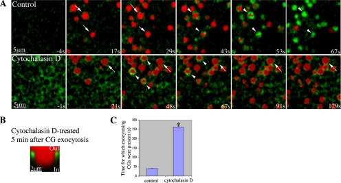
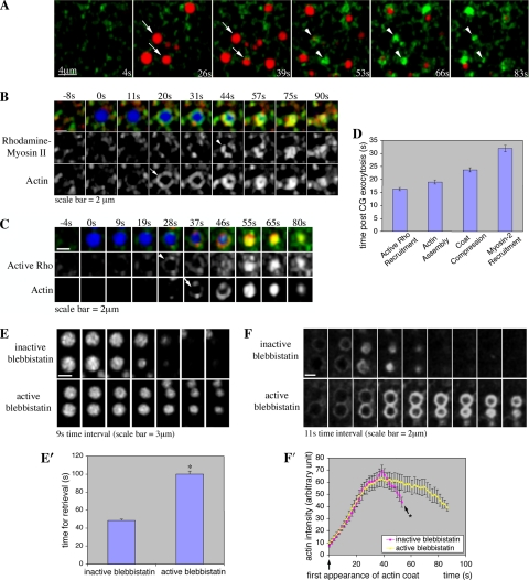
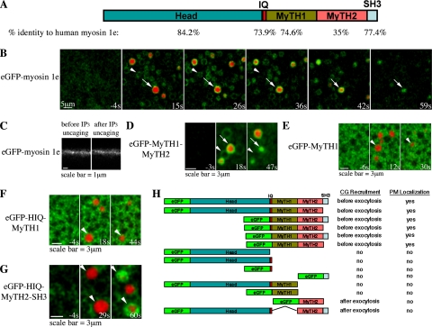
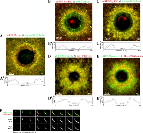
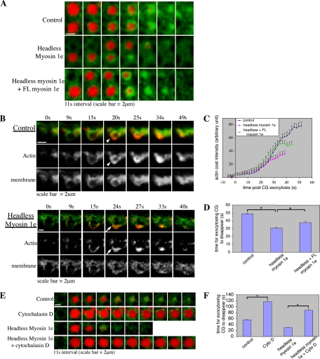
Actin is involved in endocytosis in organisms ranging from yeast to mammals. In activated Xenopus eggs, exocytosing cortical granules (CGs) are surrounded by actin "coats," which compress the exocytosing compartments, resulting in compensatory endocytosis. Here, we examined the roles of two myosins in actin coat compression. Myosin-2 is recruited to exocytosing CGs late in coat compression. Inhibition of myosin-2 slows coat compression without affecting actin assembly. This differs from phenotype induced by inhibition of actin assembly, where exocytosing CGs are trapped at the plasma membrane (PM) completely. Thus, coat compression is likely driven in part by actin assembly itself, but it requires myosin-2 for efficient completion. In contrast to myosin-2, the long-tailed myosin-1e is recruited to exocytosing CGs immediately after egg activation. Perturbation of myosin-1e results in partial actin coat assembly and induces CG collapse into the PM. Intriguingly, simultaneous inhibition of actin assembly and myosin-1e prevents CG collapse. Together, the results show that myosin-1e and myosin-2 are part of an intricate machinery that coordinates coat compression at exocytosing CGs.


Figures

Figure 1.
Figure 1.
Cytochalasin D traps exocytosing CGs at the PM. (A) Subcortical view showing actin coats (Alexa488-G-actin, arrowhead) assemble around exocytosing CGs after CG exocytosis (TR-dextran, arrow) in control cells. On treatment with cytochalasin D, exocytosing CGs (TR-dextran) remains present without being compressed for extended period. In the presence of cytochalasin D, most CGs (arrows) fail to form coats after exocytosis, although one or two do form coats (arrowheads) but then fail to compress. IP3 uncaging occurred at 0s. (B) Z-view showing that CG compartment (revealed by TR-dextran) remains trapped at the PM 5 min after exocytosis when treated with cytochalasin D. (C) Quantification of time for which the exocytosing CGs are present (n = 84 for control, n = 69 for cytochalasin D treated; asterisk indicates a p = 1 × 10−114; error bar is SEM). For control, the time represents the average time taken to retrieve the exocytosing CG membrane. For cytochalasin D-treated cells, the time represents a significant underestimation because most CGs are still present after 4.5 min.
Figure 2.
Figure 2.
Myosin-2 is required for efficient coat compression. (A) Subcortical view showing myosin-2 (rhodamine myosin-2, pseudocolored in green, arrowhead) is recruited to exocytosing CGs (Alexa647 dextran, pseudocolored in red, arrow) after onset of coat compression. IP3 uncaging occurred at 0s. (B) Subcortical view showing simultaneous imaging of myosin-2 and actin recruitment to CG in the presence of Alexa647 dextran (blue). Myosin-2 (rhodamine myosin-2, red, arrowhead) is recruited to exocytosing CGs (Alexa647 dextran) after actin coat (Alexa488 G-actin, green, arrow) is assembled. 0s indicates time at which CG exocytoses. (C) Subcortical view showing simultaneous imaging of active RhoA and actin recruitment to CG in the presence of Alexa647 dextran (blue). RhoA is activated (rGBD-eGFP, green, arrowhead) at the exocytosing CGs (Alexa647 dextran) just before the onset of actin coat (Utr1-261-mRFP, red, arrow) assembly. 0s indicates the time at which CG exocytoses. (D) Time at which myosin-2 and active RhoA are recruited to CGs with respect to the onset of actin assembly and coat compression (all of which are statistically different from one another with p < 0.001). (E) Subcortical view showing exocytosing CGs (TR-dextran) take longer to be retrieved when treated with active blebbistatin. The first frame indicates the time at which CGs first exocytosed. (E′) Quantification of time taken to retrieve CG membrane [n = 68 for inactive (+)-blebbistatin, n = 70 for active (±)-blebbistatin; error bar is SEM; asterisk indicates a p = 1 × 10⁻²⁷]. (F) Subcortical view showing that active (±)-blebbistatin prolongs the presence of actin coats, without affecting the rate of actin coat (Utr1-261-mRFP) assembly compared with inactive (+)-blebbistatin–treated cells. The first frame indicates the first appearance of actin coat. (F′) Quantification of the intensity of actin coats at exocytosing CGs (n = 29 for each group). No data are available for time points after that noted with asterisk, because CG membrane is retrieved by then (see E and E′); consequently, there is no longer any actin coat (see F).
Figure 3.
Figure 3.
Myosin-1e is transiently recruited to all CGs upon calcium elevation, but it only remains on those that have exocytosed. (A) Schematic representation of the primary structure of X. laevis myosin-1e. The percentage of sequence identity to human myosin-1e for each domain is shown under the respective domain under the primary structure. (B) Subcortical view showing that eGFP-myosin-1e is recruited to all CGs upon calcium elevation, but that it only remains on those that have fused with PM (TR-dextran, arrow), and that it disappears from those that have not exocytosed (arrowhead). (C) Z view showing eGFP-myosin-1e is present on the PM before IP3 uncaging. (D) Subcortical view showing that eGFP-MyTH1-MyTH2 is recruited to all CGs upon calcium elevation, but it only remains on those that have fused with PM (TR-dextran, arrow), and it disappears from those that have not exocytosed (arrowhead). (E) Subcortical view showing that eGFP-MyTH1 is not recruited to exocytosing CGs (TR-dextran, arrowhead). (F) Subcortical view showing that eGFP-HIQ-MyTH1 (HIQ stands for Head and IQ domains) is not recruited to exocytosing CGs (TR-dextran, arrowhead). (G) Subcortical view showing that eGFP-HIQ-MyTH2-SH3 is recruited to CGs after exocytosis (TR-dextran, arrowhead). (H) Summary of the recruitment of different eGFP fusion proteins containing different domains of myosin-1e. IP3 uncaging occurs at 0s in B and D–G.
Figure 4.
Figure 4.
MyTH2 recruitment to CGs depends on actin coat assembly. (A) Subcortical images showing that mRFP-MyTH2 (red, arrowhead) is recruited to the exocytosing CGs (Alexa647 dextran, blue) at about the same time as the onset of actin coat (Alexa488 G-actin, green, arrow) assembly. 0s indicates the time at which CG exocytoses. (B) Quantification of the intensity of MyTH2 and actin coats on CGs (n = 10). (C) Subcortical images showing that inhibition of actin coat (Alexa488 G-actin, green) assembly by cytochalasin D also prevents mRFP-MyTH2 (red) recruitment to exocytosing CGs (Alexa647 dextran, blue). 0s indicates the time at which CG exocytoses. (D) F-actin cosedimentation assay showing that MyTH2 associates with F-actin. Oocytes microinjected with the indicated final concentration of GST or GST-MyTH2 RNA, with or without pretreatment of latrunculin, are homogenized and ultracentrifuged to pellet the F-actin. GST was present in cells injected with RNA encoding GST-MyTH2, probably due to proteolysis of the GST-MyTH2 construct. P, pellet; S, supernatant.
Figure 5.
Figure 5.
MyTH2 preferentially associates with the newly assembled actin. Compressed 4D images showing (A) the more stable F-actin (Utr1-261-mRFP, red) resides closer to the wound border compared with the newly assembled actin (Alexa488 G-actin, green), which is further illustrated in the line scan shown in A′ where signals of the Utr1-261 (red) and G-actin (green) are measured by drawing a line from one side of the wound to the other. (B) mRFP-MyTH2 (red) is localized further away from the wound border compared with the more stable F-actin (Utr1-261-eGFP, green), which is further illustrated in the line scan shown in (B′) where signals of the Utr1-261 (green) and MyTH2 (red) are measured by drawing a line from one side of the wound to the other. (C) mRFP-MyTH2 (red) colocalizes with the newly assembled actin (Alexa488 G-actin, green) around the oocyte wound; which is further illustrated in the line scan shown in C′ where signals of the G-actin (green) and MyTH2 (red) are measured by drawing a line from one side of the wound to the other. (D) eGFP-myosin-1e (green) localizes with the stable F-actin (Utr1-261-mRFP, red) around the oocyte wound, which is further illustrated in the line scan shown in D′ where signals of the Utr1-261 (red) and myosin-1e (green) are measured by drawing a line from one side of the wound to the other. (E) eGFP-myosin-1e (green) localizes with the newly assembled actin (Alexa568 G-actin, red) around the oocyte wound, which is further illustrated in the line scan shown in E′ where signals of the myosin-1e (green) and G-actin (red) are measured by drawing a line from one side of the wound to the other. Oocytes are wounded at 0s in A–E. (F) Images showing the assembly of a newly polymerized actin comet, where mRFP-MyTH2 (red) and Utr1-261-eGFP (green) are present. Notice that mRFP-MyTH2 (red) is more prominent at the head (arrow), rather than the tail (arrowhead) of the actin comet.
Figure 6.
Figure 6.
Myosin-1e prevents premature coat compression and ensures symmetric coat assembly. (A) Subcortical images showing expression of the headless myosin-1e leads to faster disappearance of exocytosing CGs (TR-dextran) compare with control. Expression of FL (full-length) myosin-1e in cells expressing the headless myosin-1e rescues the phenotype induced by the headless myosin-1e. The first frame indicates the time at which CGs exocytosed. (B) Z views showing PM (farnesylated-eGFP, green) incorporates into the CG membrane upon CG exocytosis, similar to that illustrated in Yu and Bement (2007). In control cells, the exocytosing CG is subsequently encircled by actin coat (Utr1-261-mRFP, red, arrowhead). Headless myosin-1e leads to incomplete coat assembly where actin (Utr1-261-mRFP, red, arrow) is assembled mostly at the basal side of the exocytosing CG (arrow). The first frame indicates the time at which CG exocytosed. (C) Quantification of actin coat intensity, as measured from subcortical images similar to that in A. Actin assembly in cells expressing headless myosin-1e is significantly less than control ∼30s after CG exocytosis (p < 0.01). Coexpression of FL myosin-1e and headless myosin-1e rescues the phenotype. (D) Quantification of time taken for the exocytosing CGs to disappear when imaged subcortically (n = 80 for control; n = 86 for headless myosin-1e; n = 70 for headless myosin-1e with FL myosin-1e; p = 1 × 10⁻¹⁸ for control vs. headless myosin; p = 0.0005 for headless myosin-1e vs. coexpression with FL myosin-1e). (E) Subcortical images showing that cytochalasin D treatment in headless myosin-1e-expressing cells results in similar phenotype as cytochalasin D treatment in nonheadless myosin-1e-expressing cells. (F) Quantification of time taken for the exocytosing CGs to disappear when imaged subcortically (n = 95 for control; n = 67 for cytochalasin D [Cyto D] treated; n = 79 for headless myosin-1e; n = 70 for headless myosin-1e with cytochalasin D treatment; asterisks represent p < 5 × 10⁻⁶). Note that the time shown for cytochalasin D-treated cells (in both normal cells and headless myosin-1e-expressing cells) is an underestimation, because some CGs are still present after the data collection time.

Similar articles

Cited by

References

    1. Becker K. A., Hart N. H. Reorganization of filamentous actin and myosin-II in zebrafish eggs correlates temporally and spatially with cortical granule exocytosis. J. Cell Sci. 1999;112:97–110. - PubMed
    1. Benink H. A., Bement W. M. Concentric zones of active RhoA and Cdc42 around single cell wounds. J. Cell Biol. 2005;168:429–439. - PMC - PubMed
    1. Bose A., Guilherme A., Robida S. I., Nicoloro S. M., Zhou Q. L., Jiang Z. Y., Pomerleau D. P., Czech M. P. Glucose transporter recycling in response to insulin is facilitated by myosin Myo1c. Nature. 2002;420:821–824. - PubMed
    1. Burkel B. M., von Dassow G., Bement W. M. Versatile fluorescent probes for actin filaments based on the actin-binding domain of utrophin. Cell Motil. Cytoskeleton. 2007 (in press). Epub 8 August 2007. - PMC - PubMed
    1. Doberstein S. K., Pollard T. D. Localization and specificity of the phospholipid and actin binding sites on the tail of Acanthamoeba myosin IC. J. Cell Biol. 1992;117:1241–1249. - PMC - PubMed

Publication types

LinkOut - more resources