. 2007 Oct;18(10):4190-9.
doi: 10.1091/mbc.e06-07-0659. Epub 2007 Aug 15.

Nuclear localization of the ERK MAP kinase mediated by Drosophila alphaPS2betaPS integrin and importin-7

Affiliations

Nuclear localization of the ERK MAP kinase mediated by Drosophila alphaPS2betaPS integrin and importin-7

Brian P James et al. Mol Biol Cell. 2007 Oct.

Abstract

The control of gene expression by the mitogen-activated protein (MAP) kinase extracellular signal-regulated kinase (ERK) requires its translocation into the nucleus. In Drosophila S2 cells nuclear accumulation of diphospho-ERK (dpERK) is greatly reduced by interfering double-stranded RNA against Drosophila importin-7 (DIM-7) or by the expression of integrin mutants, either during active cell spreading or after stimulation by insulin. In both cases, total ERK phosphorylation (on Westerns) is not significantly affected, and ERK accumulates in a perinuclear ring. Tyrosine phosphorylation of DIM-7 is reduced in cells expressing integrin mutants, indicating a mechanistic link between these components. DIM-7 and integrins localize to the same actin-containing peripheral regions in spreading cells, but DIM-7 is not concentrated in paxillin-positive focal contacts or stable focal adhesions. The Corkscrew (SHP-2) tyrosine phosphatase binds DIM-7, and Corkscrew is required for the cortical localization of DIM-7. These data suggest a model in which ERK phosphorylation must be spatially coupled to integrin-mediated DIM-7 activation to make a complex that can be imported efficiently. Moreover, dpERK nuclear import can be restored in DIM-7-deficient cells by Xenopus Importin-7, demonstrating that ERK import is an evolutionarily conserved function of this protein.


Figures
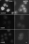
Figure 1.
Figure 1.
DIM-7 is important for nuclear dpERK accumulation in spreading S2 cells. Integrin expressing, spreading cells were treated with control dsRNA (against the endogenous βPS) or one of two nonoverlapping dsRNAs that target DIM-7 (mskA and mskB, middle and bottom rows, respectively). The cells were fixed and stained with antibodies against DIM-7 (left column) or dpERK (right column). The DIM-7 dsRNAs reduce both DIM-7 and nuclear dpERK; mskA is slightly more effective in each assay, and was used in subsequent experiments. Bar, 10 μm.
Figure 2.
Figure 2.
DIM-7 is important for nuclear dpERK. (A) Spreading S2 cells expressing αPS2βPS integrins were stained for dpERK 30 min after plating on glass coverslips coated with RBB-Tiggrin. Nuclear dpERK was quantitated microscopically in control cells (treated with dsRNA against endogenous βPS) or cells treated with DIM-7 (mskA) dsRNA (control value normalized to 1). (B) Similar to (A), except cells were plated and serum starved overnight, and assayed 20 min after stimulation with insulin. Transfection with Xenopus Importin-7 largely rescues the inhibition of nuclear dpERK seen with dsRNA against DIM-7. In all cases, only cells that spread to similar extents were measured (see text). Error bars indicate standard deviations from averages for three experiments; each experiment represents a minimum of 10 cells from seven different fields. Representative images of treated cells can be found in Supplemental Figure 3.
Figure 3.
Figure 3.
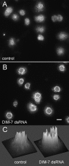
Distribution of total ERK in integrin-expressing, spreading S2 cells changes after DIM-7 reduction. αPS2βPS integrin-transfected cells were stained for total ERK (compared with dpERK staining in similarly treated cells in Figure 1). Cells in A were treated with only control dsRNA against endogenous βPS; cells in B were also grown in dsRNA targeting DIM-7. Most cytoplasmic ERK is present in aggregates. On reduction of DIM-7, the aggregates are brighter and concentrated around the nucleus, often creating a bright perinuclear ring. This is especially evident in intensity plots of individual cells, as illustrated in C. Similar plots for 8 cells from each panel can be found in the Supplemental Figure 6. Bar, 10 μm.
Figure 4.
Figure 4.
Integrin mutations inhibit dpERK nuclear localization. Spreading (A) or insulin-stimulated (B) cells were stained for dpERK as described in Figure 2. Cells expressing mutations in integrin β subunits that alter cytoplasmic residues (G1) or ligand binding (G4) greatly reduce nuclear dpERK, even though these cells are spread to similar extents. (The βPS-G4 cells are spread independently of their integrins; see text.) Error bars indicate standard deviations from averages from three experiments; each experiment represents a minimum of 10 cells from seven different fields. Representative images of treated cells can be found in Supplemental Figure 3.
Figure 5.
Figure 5.
Total phospho-ERK levels are similar in integrin mutants. Western blot showing levels of total dpERK 20 min after insulin stimulation (except lane 1) in cells expressing wild-type or mutant forms of βPS. Loading control is the same blot probed with antibodies against β-tubulin. As indicated by the quantitation below the bands (which reflects a ratio of dpERK to tubulin), dpERK is sometimes reduced in integrin mutants relative to cells expressing wild-type integrins, but integrin mutations are never seen to reduce total dpERK levels similarly to the reduction in nuclear dpERK at the same time point (Figure 4).
Figure 6.
Figure 6.
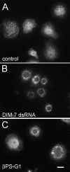
Total ERK in insulin treated, integrin expressing cells. As for spreading cells (Figure 3), total cytoplasmic ERK in control cells (expressing wild-type αPS2βPS) is found largely in aggregates (A), which tend to localize around the nucleus in cells treated with dsRNA against DIM-7 (B). Cells that express the cytoplasmic βPS-G1 mutant integrin with αPS2 (C) are more variable; most show similarities to DIM–depleted cells, although some, such as the cell on the left, look much like control cells. All cells are treated with dsRNA against the endogenous βPS gene. Bar, 10 μm.
Figure 7.
Figure 7.
DIM-7 tyrosine phosphorylation is affected by integrins. Proteins were immunoprecipitated by anti-DIM-7 from cells 10 min after stimulation by insulin (or after no insulin, lane 1). The cell lines used were transformed to express no integrin or αPS2 in combination with the βPS subunit indicated. All cells (including controls) were grown in dsRNA to inhibit expression of endogenous βPS. Expression of the mutant βPS subunits does not significantly alter the amount of DIM-7 that is brought down, but it does reduce its tyrosine phosphorylation.
Figure 8.
Figure 8.
DIM-7 and integrin colocalization. Drosophila S2 cells expressing αPS2βPS integrins were spread on a fragment of the ECM ligand Tiggrin, and fixed and stained to show the distributions of various components. (A and B) DIM-7 is found at high levels around the cell nucleus, and it also is localized at the cell periphery where concentrations of integrins (stained with anti-βPS) are mediating cell spreading (arrows). (C and D) DIM-7 is specifically absent from focal adhesions (visualized by high-density accumulations of paxillin, arrows) and the surrounding areas of the cell periphery. More diffuse paxillin and DIM-7 are seen along spreading edges of the cell (e.g., asterisks). Bar, 10 μm.
Figure 9.
Figure 9.
DIM-7 colocalizes with integrins but not with organized adhesive sites. Confocal images of peripheral areas of cells plated on RBB-Tiggrin, as in Figure 8, but at higher resolution. As shown in the top row, DIM-7 and integrins are largely concentrated in the same regions near the edge of the cell. However, where more organized adhesive sites are formed (shown by paxillin staining, lower row), there is no concomitant concentration of DIM-7. Bar, 5 μm.
Figure 10.
Figure 10.
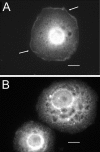
Peripheral localization of DIM-7 is Corkscrew dependent. (A) Integrin-expressing cells were spread on RBB-Tiggrin and stained for DIM-7. Note the DIM-7 staining around virtually the entire periphery in this cell (arrows). (B) DIM-7 peripheral localization is absent in cells that have been treated with dsRNA against corkscrew, although DIM-7 staining seems greater overall than in A; Western blots confirm that DIM-7 levels increase in cells with reduced Corkscrew (data not shown). Bar, 10 μm.
Figure 11.
Figure 11.
Model for the role of integrins in ERK nuclear import in S2 cells. DIM-7 is localized near functioning integrins, at least in part by its association with the SHP-2 phosphatase Corkscrew. dpERK (activated via either hormone or cell spreading) must associate with activated (perhaps by tyrosine phosphorylation) DIM-7 at the periphery, after which the ERK/DIM-7 complex is imported into the nucleus. In cells expressing mutant integrins, ERK can be phosphorylated and transported centrally, but this ERK cannot efficiently cross the nuclear envelope, even though there is a high perinuclear concentration of DIM-7. It is not known whether this is because the ERK associates with an inhibitor in the absence of activated DIM-7 at the periphery or whether the perinuclear DIM-7 is not activated in the same way as the cortical importin. Note that DIM-7 is not required for nuclear directed transport, only nuclear import, because the perinuclear ERK aggregates are formed in DIM-7–depleted cells as well as those expressing mutant integrins.

Similar articles

Cited by

References

    1. Adachi M., Fukuda M., Nishida E. Two co-existing mechanisms for nuclear import of MAP kinase: passive diffusion of a monomer and active transport of a dimer. EMBO J. 1999;18:5347–5358. - PMC - PubMed
    1. Allard J. D., Chang H. C., Herbst R., McNeill H., Simon M. A. The SH2-containing tyrosine phosphatase corkscrew is required during signaling by sevenless, Ras1 and Raf. Development. 1996;122:1137–1146. - PubMed
    1. Allard J. D., Herbst R., Carroll P. M., Simon M. A. Mutational analysis of the SRC homology 2 domain protein-tyrosine phosphatase Corkscrew. J. Biol. Chem. 1998;273:13129–13135. - PubMed
    1. Aplin A. E., Stewart S. A., Assoian R. K., Juliano R. L. Integrin-mediated adhesion regulates ERK nuclear translocation and phosphorylation of Elk-1. J. Cell Biol. 2001;153:273–282. - PMC - PubMed
    1. Assoian R. K., Schwartz M. A. Coordinate signaling by integrins and receptor tyrosine kinases in the regulation of G1 phase cell-cycle progression. Curr. Opin. Genet. Dev. 2001;11:48–53. - PubMed

Publication types

MeSH terms

LinkOut - more resources