. 2007 Aug 21;104(34):13678-83.
doi: 10.1073/pnas.0703650104. Epub 2007 Aug 15.

Dissociation of the insulin receptor and caveolin-1 complex by ganglioside GM3 in the state of insulin resistance

Affiliations

Dissociation of the insulin receptor and caveolin-1 complex by ganglioside GM3 in the state of insulin resistance

Kazuya Kabayama et al. Proc Natl Acad Sci U S A. .

Abstract

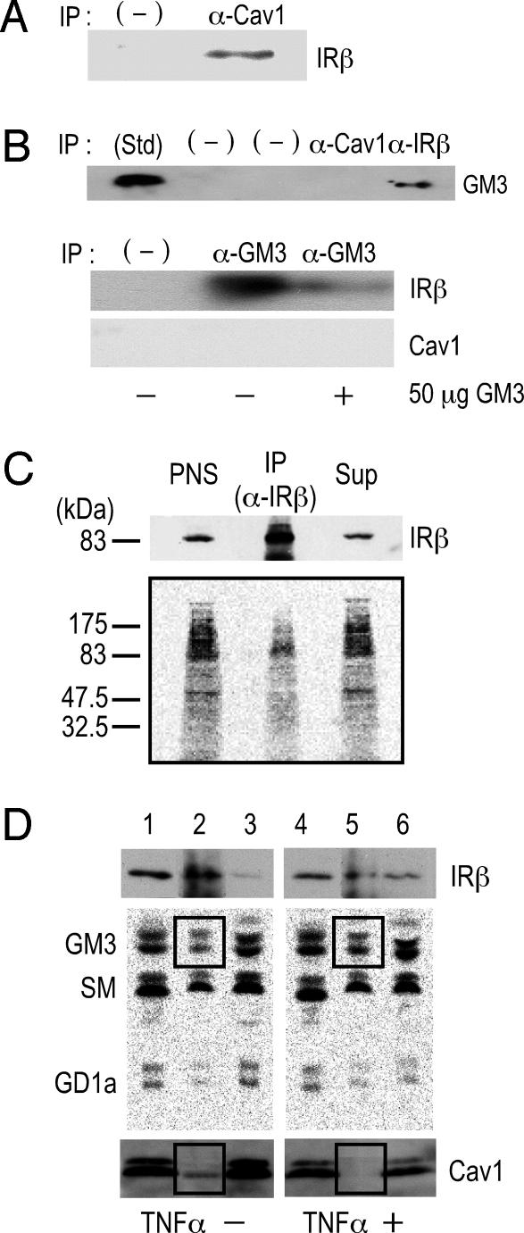
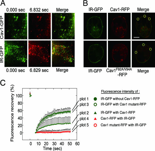
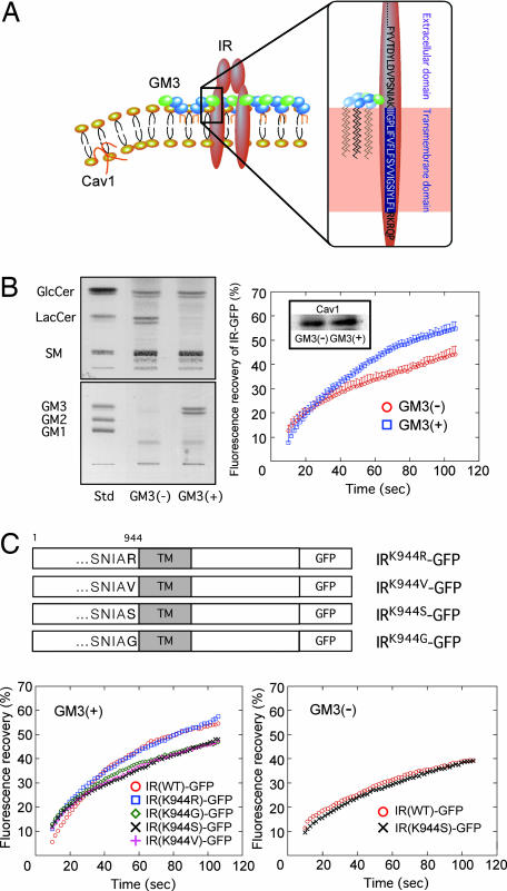
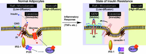
Membrane microdomains (lipid rafts) are now recognized as critical for proper compartmentalization of insulin signaling. We previously demonstrated that, in adipocytes in a state of TNFalpha-induced insulin resistance, the inhibition of insulin metabolic signaling and the elimination of insulin receptors (IR) from the caveolae microdomains were associated with an accumulation of the ganglioside GM3. To gain insight into molecular mechanisms behind interactions of IR, caveolin-1 (Cav1), and GM3 in adipocytes, we have performed immunoprecipitations, cross-linking studies of IR and GM3, and live cell studies using total internal reflection fluorescence microscopy and fluorescence recovery after photobleaching techniques. We found that (i) IR form complexes with Cav1 and GM3 independently; (ii) in GM3-enriched membranes the mobility of IR is increased by dissociation of the IR-Cav1 interaction; and (iii) the lysine residue localized just above the transmembrane domain of the IR beta-subunit is essential for the interaction of IR with GM3. Because insulin metabolic signal transduction in adipocytes is known to be critically dependent on caveolae, we propose a pathological feature of insulin resistance in adipocytes caused by dissociation of the IR-Cav1 complex by the interactions of IR with GM3 in microdomains.


Conflict of interest statement

The authors declare no conflict of interest.

Figures

Fig. 1.
Fig. 1.
The IR forms distinct complexes with Cav1 and GM3 in 3T3-L1 adipocytes. (A) Interaction of Cav1 and IR. PNS of whole-cell lysates were immunoprecipitated with an anti-Cav1 antibody or anti-mouse IgG (−), and the precipitates were subjected to SDS/PAGE followed by immunoblotting with an anti-IRβ antibody. (B) GM3 associates with IR but not with Cav1. (Upper) PNS were immunoprecipitated with an anti-Cav1 antibody, an anti-IRβ antibody, or an negative control antibody (−). The precipitates were subjected to TLC followed by immunostaining with the anti-GM3 antibody M2590 as described in Materials and Methods. (Lower) Immunoprecipitation was performed with the anti-GM3 antibody DH2, in the presence or absence of 50 μg of GM3 or with anti-mouse IgG (−). The precipitates were then subjected to SDS/PAGE, followed by immunoblotting with an anti-IRβ or anti-Cav1 antibody. (C) GM3 directly binds to IR. A cross-linking assay of GM3 and IR in adipocytes was performed by using photoactivatable 3H-labeled GM3 described in Materials and Methods. After cross-linking, cells were then lysed and subjected to immunoprecipitation with an anti-IRβ antibody. PNS, anti-IRβ immunoprecipitates (IP), and the supernatant from the immunoprecipitation (Sup) were subjected to SDS/PAGE followed by immunoblotting with an anti-IRβ antibody and autoradiography. (D) Formation of the IR–GM3 complex is increased relative to that of the IR–Cav1 complex in adipocytes in a state of TNFα-induced insulin resistance. Glycosphingolipids in adipocytes were metabolically labeled with [3H]sphingosine, and the cells were left untreated or treated with 0.1 nM TNFα. A coimmunoprecipitation assay was performed on cell lysates by using an anti-IRβ antibody. Samples (equivalent in radioactivity) of PNS, anti-IRβ immunoprecipitate, and the corresponding supernatant, obtained from cells untreated (lanes 1–3) and treated with TNFα (lanes 4–6), were subjected to SDS/PAGE followed by immunoblotting with an anti-IRβ antibody (Top) or an anti-Cav1 antibody (Bottom). Radioactive lipids were extracted and separated by HPTLC and visualized by autoradiography. In all experiments, reproducible results were obtained, and representative data are presented.
Fig. 2.
Fig. 2.
Immobilization of IR by Cav1 in living cells. (A) TIR-FM analyses. Cav1-GFP and IR-GFP were expressed in HEK293 cells, and time-lapse images at the cell surface were taken (SI Movie 1 for Cav1-GFP and SI Movie 2 for IR-GFP). Selected frames in the same area between 0 sec (green) and 7 sec (pseudored) and their merged images are shown. (B) FRAP analyses. Cav1-RFP or Cav1F92A/F94A-RFP was coexpressed with IR-GFP in HEK293 cells. The area at the cell surface of aggregated Cav-1 or mutant protein was identified by confocal images (Upper), and the areas were bleached. (C) The fluorescence recovery of IR-GFP expressed alone (plot 1) or coexpressed with Cav1-RFP (plot 2) or the Cav1-RFP mutant (plot 3), and the fluorescence recovery of Cav1-RFP (plot 4) or the Cav1-RFP mutant (plot 5) coexpressed with IR-GFP were measured.
Fig. 3.
Fig. 3.
The lysine residue IR944 is essential for the interaction of IR with GM3. (A) Schematic representation of the proposed interaction of a lysine residue at IR944, which is located just above the transmembrane domain, and GM3 at the cell surface. (B) Enhanced mobility of IR in GM3-enriched membrane. (Left) Glycosphingolipid analysis of GM3-reconstituted cells [GM3 (+)] and mock cells [GM3 (−)]. Glycosphingolipids extracted from these cells, corresponding to 1 mg of cellular protein, were separated on HPTLC plates and stained with resorcinol–HCl reagent to visualize gangliosides, or with cupric acetate–phosphoric acid reagent for neutral glycosphingolipids as described in Materials and Methods. (Right) FRAP analyses. Shown is fluorescence recovery of IR-GFP in GM3 (−) and GM3 (+) cells expressing equal levels of Cav1 (Inset). (C) Specificity of the interaction between lysine at IR944 and GM3 by FRAP analyses. (Upper) Schematic structure of IR-GFP mutants in which the lysine at IR944 is replaced with basic and neutral amino acids. (Lower Left) Fluorescence recovery of IR-GFP mutants in GM3 (+) cells. (Lower Right) Fluorescence recovery of IR-GFP mutants in GM3 (−) cells.
Fig. 4.
Fig. 4.
Proposed mechanism behind the shift of IR from the caveolae to the GEMs in adipocytes during a state of insulin resistance. Shown is a schematic representation of raft/microdomains comprising caveolae and noncaveolae rafts such as GEM. Caveolae and GEM reportedly can be separated by an anti-Cav1 antibody (14). IR may be constitutively resident in caveolae via its binding to the scaffolding domain of Cav1 through the caveolin binding domain in its cytoplasmic region. Binding of IR and Cav1 is necessary for successful insulin metabolic signaling (Table 1). In adipocytes the localization of IR in the caveolae is interrupted by elevated levels of the endogenous ganglioside GM3 during a state of insulin resistance induced by TNFα (12). The present study has proven a mechanism, at least in part, in which the dissociation of the IR–Cav1 complex is caused by the interaction of a lysine residue at IR944, located just above the transmembrane domain, and the increased GM3 clustered at the cell surface.

References

    1. Nojiri H, Stroud M, Hakomori S. J Biol Chem. 1991;266:4531–4537. - PubMed
    1. Tagami S, Inokuchi J-i, Kabayama K, Yoshimura H, Kitamura F, Uemura S, Ogawa C, Ishii A, Saito M, Ohtsuka Y, et al. J Biol Chem. 2002;277:3085–3092. - PubMed
    1. Razani B, Woodman SE, Lisanti MP. Pharmacol Rev. 2002;54:431–467. - PubMed
    1. Goldberg RI, Smith RM, Jarett L. J Cell Physiol. 1987;133:203–212. - PubMed
    1. Gustavsson J, Parpal S, Karlsson M, Ramsing C, Thorn H, Borg M, Lindroth M, Peterson KH, Magnusson KE, Stralfors P. FASEB J. 1999;13:1961–1971. - PubMed

Publication types