. 2007 Aug 15;27(33):8745-56.
doi: 10.1523/JNEUROSCI.1002-07.2007.

Calcineurin in reactive astrocytes plays a key role in the interplay between proinflammatory and anti-inflammatory signals

Affiliations

Calcineurin in reactive astrocytes plays a key role in the interplay between proinflammatory and anti-inflammatory signals

Ana M Fernandez et al. J Neurosci. .

Abstract

Maladaptive inflammation is a major suspect in progressive neurodegeneration, but the underlying mechanisms are difficult to envisage in part because reactive glial cells at lesion sites secrete both proinflammatory and anti-inflammatory mediators. We now report that astrocytes modulate neuronal resilience to inflammatory insults through the phosphatase calcineurin. In quiescent astrocytes, inflammatory mediators such as tumor necrosis factor-alpha (TNF-alpha) recruits calcineurin to stimulate a canonical inflammatory pathway involving the transcription factors nuclear factor kappaB (NFkappaB) and nuclear factor of activated T-cells (NFAT). However, in reactive astrocytes, local anti-inflammatory mediators such as insulin-like growth factor I also recruit calcineurin but, in this case, to inhibit NFkappaB/NFAT. Proof of concept experiments in vitro showed that expression of constitutively active calcineurin in astrocytes abrogated the inflammatory response after TNF-alpha or endotoxins and markedly enhanced neuronal survival. Furthermore, regulated expression of constitutively active calcineurin in astrocytes markedly reduced inflammatory injury in transgenic mice, in a calcineurin-dependent manner. These results suggest that calcineurin forms part of a molecular pathway whereby reactive astrocytes determine the outcome of the neuroinflammatory process by directing it toward either its resolution or its progression.


Figures
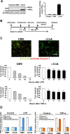
Figure 1.
Figure 1.
Astrocytic calcineurin and neuroinflammatory damage. A, Astrocytes transduced with ΔCnA show increased activity of calcineurin (measured as release of PO4) compared with mock-transfected (CMV) or wt astrocytes (histograms). Only ΔCnA-transfected astrocytes produced the truncated mutant form of calcineurin (ΔCnA; blots). Levels of endogenous calcineurin remained unaffected. B, Schedule followed for coculture of astrocytes and neurons. Astrocytes were plated and transfected with corresponding DNAs, and neurons were added 24 h later. Thereafter, cocultures were challenged with inflammatory stimuli (LPS or TNF-α) for various times before analysis of neuronal death after a total of 4 d in coculture. Inhibitors such as CsA or MG-132 were added at indicated times. C, Photomicrographs, Representative double immunocytochemical staining used to identify apoptotic (activated caspase 3+ cells; red) neurons (β3-tubulin+ cells; green) after inflammatory challenge. Histograms, Neurons cocultured with mock-transfected astrocytes (CMV; striped left histograms) die soon after LPS (top histograms) or TNF-α addition (bottom histograms), whereas when cocultured with astrocytes expressing ΔCnA (black right histograms), they show a significantly greater resistance to these inflammatory stimuli (**p < 0.01 vs respective CMV times). Number of living neurons are expressed as percentage of unstimulated control cultures at time 0. Scale bar, 100 μm. D, The release of ROS in response to proinflammatory stimuli (LPS or TNF-α) was fully abrogated when astrocytes (but not neurons) expressed ΔCnA but not the empty vector (CMV). Note that, with the doses of LPS or TNF-α used, astrocytes did not produce ROS. All experiments were repeated six times. ***p < 0.001.
Figure 2.
Figure 2.
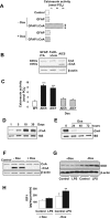
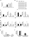
Regulated expression of ΔCnA in astrocytes. A, Coexpression of GFAP–tTA (GFAP) and TetO–ΔCnA (ΔCnA) in cultured wt astrocytes allows Dox-regulated activation of calcineurin. Both control (wt) and GFAP–tTA-transduced astrocytes show lower and Dox-independent calcineurin activity. ***p < 0.001. B, Astrocyte inducible calcineurin (AIC mouse 5) but not single mutant TetO–ΔCnA or GFAP–tTA mice produced ΔCnA protein, a truncated form of the wt protein. Wild-type calcineurin (CnA) was detected in the three animals. C, Calcineurin activity in brain extracts from AIC mice (lines 5, 7) was markedly elevated compared with a wt control and was regulated by Dox. ***p < 0.001. D, Expression of ΔCnA mRNA in the brain of AIC mice was found postnatally and maintained thereafter. E, ΔCnA mRNA levels in the brain of AIC mice were inhibited within hours after addition of Dox to the drinking water. F, Expression of ΔCnA protein in astrocytes obtained from AIC mice was regulated by Dox in the culture medium. Controls are astrocytes from wt mice. G, Astrocytes from AIC mice secrete Cu/Zn SOD (SOD) to the medium after LPS challenge in a calcineurin-dependent manner. In the presence of Dox in the culture medium (which suppresses ΔCnA expression), levels of SOD remain unaffected. Note that expression of ΔCnA is sufficient to increase SOD levels. Representative blot is shown (n = 6). H, AIC astrocytes also secrete greater amounts of IGF-I after LPS in the absence of Dox in the culture medium (*p < 0.05 vs control and **p < 0.01 vs +Dox and vs control; n = 6).
Figure 3.
Figure 3.
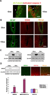
Astrocytic calcineurin protects against inflammatory damage after brain trauma. A, In AIC mice expressing ΔCnA (not treated with Dox), the number of dying neurons (β3-tubulin+-activated caspase 3+ cells) 5 d after the lesion in the area surrounding a traumatic injury (asterisk) of the parietal cortex was reduced compared with AIC mice treated with Dox for 1 week before lesion. Scale bar, 50 μm. B, Expression of ΔCnA in AIC mice (−Dox) reduced the number of reactive astrocytes (GFAP+; green) expressing Cox2 (red) in the lesioned area (asterisk). Note expression of Cox2 also by unidentified GFAP cells surrounding the lesion site. Scale bar, 100 μm. C, Levels of both Cox2 and iNOS2 at the injury site were also dramatically reduced in ΔCnA-expressing AIC mice (p < 0.001 vs AIC +Dox, for both markers; n = 10 at 5 d after lesion). Control, Sham operated; Inj, brain injury. D, Levels of MHCII (a marker of microglia activation) were increased at the lesion site 5 d after brain trauma in AIC mice not expressing ΔCnA (+Dox; n = 10), but the increase was smaller when expressing ΔCnA (−Dox; n = 10). Protein load in gels was normalized by measuring β-actin levels. Bottom histograms, Number of MHCII+, CD11b+ (a marker of infiltrating macrophages), and double MHCII+–CD11b+ cells at the lesioned site in AIC mice treated with Dox 1 week before lesion (+Dox), not treated with Dox (−Dox), or treated with Dox until 2 d after the lesion (±Dox). In the absence of Dox, when ΔCnA is expressed in astrocytes, the number of MHCII+ and MHCII+–CD11b+ cells is significantly reduced, whereas CD11b+ cells remain unaffected. Note that the reduction is also present when AIC mice started to express ΔCnA 2 d after the lesion was produced (***p < 0.001 vs −Dox and ±Dox; n = 5 per group). Photomicrographs, Representative MHCII+ and CDb11+ cells located in the vicinity of the lesion site (asterisk). Cell counts were done in double-stained brain sections. Scale bar, 50 μm.
Figure 4.
Figure 4.
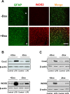
Astrocytic calcineurin protects against LPS-induced inflammatory damage. A, Three days after intraparenchymael injection of LPS, reactive astrocytes (GFAP+) in AIC mice not treated with Dox show negligible iNOS2 immunoreactivity. Scale bar, 100 μm. B, This was paralleled by a drastic reduction in levels of Cox2 and iNOS2 in the injected area (p < 0.001 vs AIC +Dox; n = 10). C, Levels of Cox2 and iNOS2 in cortical tissue of intraperitoneally LPS (iLPS)-injected AIC (−Dox) mice were reduced compared with AIC mice receiving Dox in the drinking water (p < 0.001 vs AIC +Dox; n = 10 at 3 d after injection). Representative blots are shown.
Figure 5.
Figure 5.
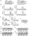
Calcineurin inhibits the NFκB/NFAT proinflammatory pathway in astrocytes. A, LPS failed to stimulate Cox2 and iNOS2 in astrocytes expressing ΔCnA. A representative blot is shown (n = 6). B, LPS- or TNF-α-induced activation of NFκB and NFAT was abrogated in ΔCnA-transduced astrocytes. The activity of these transcription factors was inhibited after exposure to LPS/TNF-α in ΔCnA-expressing astrocytes. Note that expression of ΔCnA in unstimulated astrocytes reduced the activity of NFκB and NFAT. CMV, Astrocytes transfected with the empty vector; ΔCnA, astrocytes transfected with constitutively active calcineurin; CMV+NFκB or NFAT, mock-transfected astrocytes expressing the gene-reporter system for either transcription factor; ΔCnA+NFκB or NFAT, ΔCnA-transfected astrocytes expressing the gene-reporter system. ***p < 0.001 versus respective controls (n = 6). C, In astrocytes from AIC mice, activation of NFκB and NFAT after LPS (left) or TNF-α (right) was regulated by Dox, confirming that expression of ΔCnA suppresses it. **p < 0.01 and ***p < 0.001 versus respective controls. D, Expression of ΔCnA blocks phosphorylation of IκBα induced by TNF-α (20 ng/ml, 15 min of exposure, representative blot). Mock-transfected cultures received the pCMV vector. E, Exposure of mock-transfected astrocytes to LPS elicited an increase in NFκBp65 (left) and NFAT4 (right) protein levels 16 h later. This increase was significantly attenuated in astrocytes expressing ΔCnA. F, The inhibitory effect of ΔCnA on NFκBp65/NFAT4 levels was blocked by the proteasome inhibitory drug MG-132 (MG). G, ΔCnA-expressing astrocytes have significantly increased NFκBp65 and NFAT4 complexed with ubiquitin. Immunoprecipitation with anti-NFκBp65 or anti-NFAT4 followed by blotting with anti-ubiquitin (top blots). H, Astrocytes transduced with ΔCnA had increased levels of NFκBp65/PPARγ and NFAT/GATA3 compared with control astrocytes. Immunoprecipitation with anti-PPARγ and anti-GATA3 followed by blotting with either anti-NFκBp65 or anti-NFAT4 (top blots). The opposite immunoprecipitations gave similar results (data not shown). In all cases, levels of β-actin or the respective immunoprecipitated protein were determined for protein load (bottom blots). Representative blots (of 6 for each experiment) are shown. All changes were significantly different (see Results).
Figure 6.
Figure 6.
Astrocytic calcineurin participates in proinflammatory and in anti-inflammatory signaling. A, In wt astrocytes, calcineurin activity was incremented by proinflammatory stimuli such as LPS/TNF-α and by neuroprotective signals such as IGF-I. **p < 0.01 and ***p < 0.001 versus control. B, In astrocytes transfected with a calcineurin-specific siRNA (CnA3), the production of inflammatory mediators such as iNOS2 or Cox2 in response to inflammatory challenge (LPS) was abrogated in parallel to inhibited calcineurin levels. Controls, Astrocytes transfected with non-sense siRNA; GAPDH, astrocytes transfected with GAPDH siRNA. Note that, although levels of this protein are depleted, the response to LPS/TNF-α remains intact. Representative blots are shown (n = 6). Other CnA siRNAs tested gave similar results (data not shown). C, LPS/TNF-α-stimulated activity of NFκB and NFAT was abrogated in the presence of CsA. NFκB and NFAT denote transfection of astrocytes with NFκB and NFAT reporters, respectively. **p < 0.01 and ***p < 0.001 versus untreated controls (shown in left white bars). D, IGF-I inhibited the increase induced by LPS or TNF-α in the activity of NFκB and NFAT. ***p < 0.001 versus unstimulated controls (n = 6). ***p < 0.001 versus unstimulated controls. E, IGF-I protected neurons against TNF-α-induced death only in the presence of astrocytes. This effect was blocked by CsA. Neurons were exposed to TNF-α overnight. F, Production of ROS after LPS challenge was blocked by IGF-I only in the presence of astrocytes. *p < 0.05 and ***p < 0.001 versus respective controls (n = 6).
Figure 7.
Figure 7.
Stages of the neuroinflammatory process in which astrocyte calcineurin may participate. Initiation, Inflammatory signals set in motion by the neuropathological process activate calcineurin, which in turn activate the canonical NFκB/NFAT pathway. Activation of local and peripheral proinflammatory mechanisms together with the recruitment of autocrine and paracrine neuroprotective mediators follows. The time course of this simultaneous anti-inflammatory and proinflammatory cascade may be critical to the eventual outcome of the inflammatory response. Both agonistic and antagonistic inflammatory signals are produced by reactive astrocytes and microglia, damaged neurons and activated endothelia, and eventually from peripheral cells recruited to the lesion site. Resolution, If already activated calcineurin is stimulated by signals such as IGF-I, a neuroprotective network is activated; Progression, if calcineurin continues to be activated by inflammatory signals, the inflammation proceeds and neurons die. Both phases may be reversibly interrelated depending on the time course of the pathological process. Mechanisms whereby calcineurin is recruited toward either inflammation or neuroprotection, which involve differential interactions with transcription factors such as PPARγ and GATA3 or proteasome degradation and which depend on the upstream signal stimulating calcineurin, warrant additional analysis.

Similar articles

Cited by

References

    1. Abbasi S, Lee JD, Su B, Chen X, Alcon JL, Yang J, Kellems RE, Xia Y. Protein kinase-mediated regulation of calcineurin through the phosphorylation of modulatory calcineurin-interacting protein 1. J Biol Chem. 2006;281:7717–7726. - PubMed
    1. Alfieri C, Evans-Anderson H, Yutzey K. Developmental regulation of the mouse IGF-I exon 1 promoter region by calcineurin activation of NFAT in skeletal muscle. Am J Physiol Cell Physiol. 2007;292:C1887–C1894. - PubMed
    1. Avni O, Lee D, Macian F, Szabo SJ, Glimcher LH, Rao A. T(H) cell differentiation is accompanied by dynamic changes in histone acetylation of cytokine genes. Nat Immunol. 2002;3:643–651. - PubMed
    1. Bailey ST, Ghosh S. “PPAR”ting ways with inflammation. Nat Immunol. 2005;6:966–967. - PubMed
    1. Beilharz EJ, Russo VC, Butler G, Baker NL, Connor B, Sirimanne ES, Dragunow M, Werther GA, Gluckman PD, Williams CE, Scheepens A. Co-ordinated and cellular specific induction of the components of the IGF/IGFBP axis in the rat brain following hypoxic-ischemic injury. Brain Res Mol Brain Res. 1998;59:119–134. - PubMed

Publication types

MeSH terms