. 2007 Aug 15;27(33):8790-804.
doi: 10.1523/JNEUROSCI.1847-07.2007.

Neuronal diversity in GABAergic long-range projections from the hippocampus

Affiliations

Neuronal diversity in GABAergic long-range projections from the hippocampus

Shozo Jinno et al. J Neurosci. .

Abstract

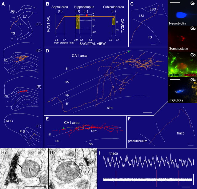
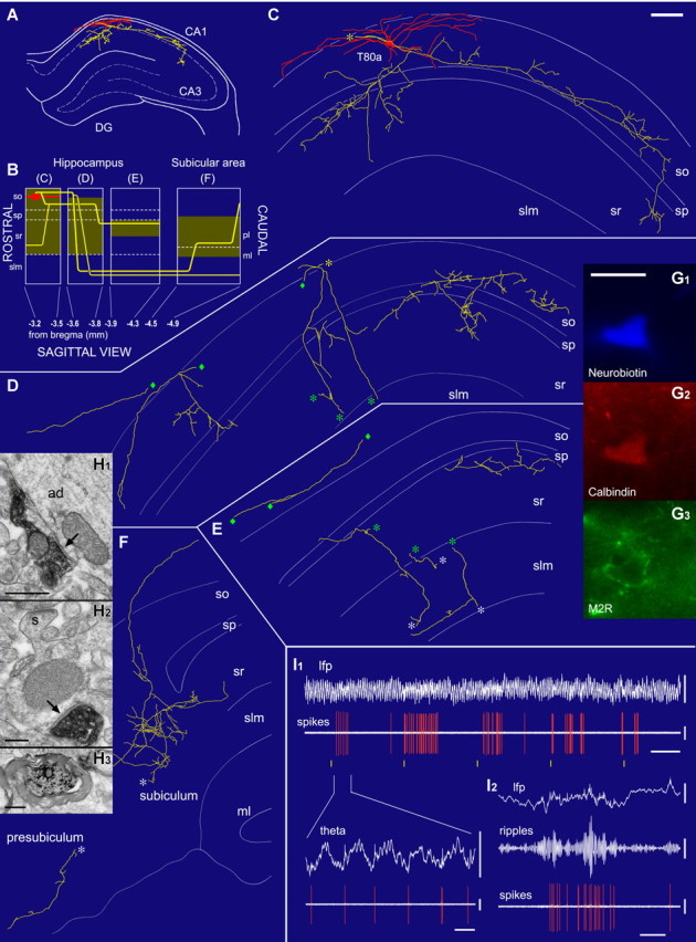
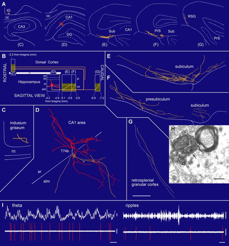
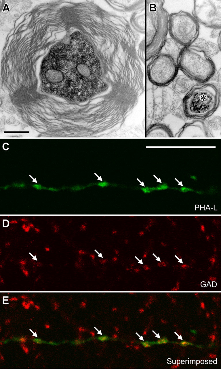
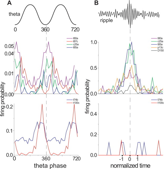
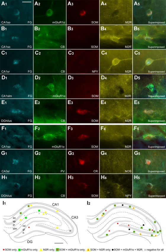
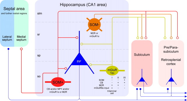
The formation and recall of sensory, motor, and cognitive representations require coordinated fast communication among multiple cortical areas. Interareal projections are mainly mediated by glutamatergic pyramidal cell projections; only few long-range GABAergic connections have been reported. Using in vivo recording and labeling of single cells and retrograde axonal tracing, we demonstrate novel long-range GABAergic projection neurons in the rat hippocampus: (1) somatostatin- and predominantly mGluR1alpha-positive neurons in stratum oriens project to the subiculum, other cortical areas, and the medial septum; (2) neurons in stratum oriens, including somatostatin-negative ones; and (3) trilaminar cells project to the subiculum and/or other cortical areas but not the septum. These three populations strongly increase their firing during sharp wave-associated ripple oscillations, communicating this network state to the septotemporal system. Finally, a large population of somatostatin-negative GABAergic cells in stratum radiatum project to the molecular layers of the subiculum, presubiculum, retrosplenial cortex, and indusium griseum and fire rhythmically at high rates during theta oscillations but do not increase their firing during ripples. The GABAergic projection axons have a larger diameter and thicker myelin sheet than those of CA1 pyramidal cells. Therefore, rhythmic IPSCs are likely to precede the arrival of excitation in cortical areas (e.g., subiculum) that receive both glutamatergic and GABAergic projections from the CA1 area. Other areas, including the retrosplenial cortex, receive only rhythmic GABAergic CA1 input. We conclude that direct GABAergic projections from the hippocampus to other cortical areas and the septum contribute to coordinating oscillatory timing across structures.


Figures

Figure 1.
Figure 1.
A double-projection neuron (T87c) recorded in vivo innervating both the septal and retrohippocampal areas. A, Representation of coronal planes showing the dendritic (red) and axonal (orange) arbors in selected sections indicated as blocks in B. B, Sagittal plane scheme of the rostrocaudal extent of the dendrites and the axon. The soma and dendrites are in stratum oriens. The axon bifurcates into a rostral (toward the septum) and a caudal (toward the subicular area) branch. Shaded areas represent axon collaterals with synaptic boutons. C–F, Reconstructions of the soma and dendrites are shown complete; the axon is shown from selected series of sections as indicated in B. The rostral axon enters the triangular septum (TS) through the lateral septum (LSI and LSD) but could not be traced to its termination. The local axon mainly innervates strata oriens (so) and radiatum (sr) but avoids str. lacunosum-moleculare (slm). The caudal axon branch enters the presubiculum from the corpus callosum (fmcc). Diamonds mark connecting points of axonal branches. G, Immunofluorescence images of the neurobiotin-filled cell body (G1) immunopositive for somatostatin (G2), calbindin (red, G3), and mGluR1α (the plasma membrane; green, G3); a neurobiotin-filled dendrite (G4) is decorated by mGluR7a-immunopositive boutons (yellow). H, Serial electron microscopic sections of a pyramidal cell dendrite (asterisk) emitting a spine (s; H2) and receiving a type II synapse (arrow) from a neurobiotin-labeled bouton (H1). I, This cell fires (bottom, orange; filter, 0.8–5 kHz) around the trough of the theta oscillations recorded extracellularly from a second electrode in the pyramidal layer (filter; dc-220 Hz). cc, Corpus callosum; IG, indusium griseum; LV, lateral ventricle; PrS, presubiculum; RSG, granular retrosplenial cortex; sp, stratum pyramidale. Scale bars: C–F, 100 μm; G1, 20 μm; G4, 5 μm; H1, 0.5 μm. Calibration: I, 0.5 s, theta, 0.5 mV, spikes, 0.5 mV.
Figure 2.
Figure 2.
An oriens-retrohippocampal projection neuron (T80a) recorded in vivo in the dorsal CA1 hippocampal area innervating the subiculum. A, The soma (red), dendrites (red), and axon (yellow) in the coronal plane at a rostrocaudal level indicated in B. B, Schematic representation in the sagittal plane showing the rostrocaudal extent of the cell. The shaded areas represent the distribution of neurobiotin-filled boutons illustrated in C–F. C–F, The soma and dendrites are shown complete; the axon is shown from a selected section as indicated by blocks in B. The local axon mainly innervates strata oriens (so), pyramidale (sp), and radiatum (sr) but mostly avoids str. lacunosum-moleculare (slm). The long-range axon collaterals pass through str. lacunosum-moleculare, extensively branch in the pyramidal layer of the subiculum, and enter the presubiculum. Asterisks and diamonds mark connecting points of axonal branches. G, Images of the neurobiotin-filled cell body (G1) show immunopositivity for calbindin in the cytoplasm (G2) and muscarinic M2 receptor in the plasma membrane (G3). H, Electron microscopy of labeled boutons making type II synapses (arrows) onto an apical dendrite (ad; H1) and a small spiny (s) dendrite (H2) of CA1 pyramidal neurons; myelinated main axon of the cell in the subicular molecular layer (H3). I, In vivo recording shows that the cell becomes active rhythmically at 0.1 Hz (I1) during theta oscillations; yellow lines mark 10 s intervals. The cell fires around the trough of theta oscillations (I1) recorded from the same electrode. During ripple episodes, the cell strongly increases its firing rate (I2; top, filter dc-220 Hz; middle, 90–140 Hz bandpass; bottom, 0.8–5 kHz). Scale bars: (in C) C–F, 100 μm; (in G1) G1G3, 20 μm; H1, 0.5 μm; H2, 0.2 μm; H3, 0.5 μm. Calibration: I1, 4 s (inset, 0.2 s); lfp, 1 mV; theta, 1 mV; spikes, 1 mV; I2, 0.2 s; lfp, 1 mV; ripples, 0.1 mV; spikes, 1 mV.
Figure 3.
Figure 3.
A radiatum-retrohippocampal projection neuron (T74b) recorded in vivo in the dorsal CA1 area innervating the subiculum (Sub), presubiculum (PrS), retrosplenial cortex (RSG), and indusium griseum (IG). A, The soma (red), dendrites (red), and axons (yellow) in coronal planes from selected sections as indicated in B. B, Representation in the sagittal plane showing the rostrocaudal extent of the dendrites and axon. The soma is located at the border of str. radiatum and lacunosum-moleculare. The axon, traced over 5 mm, runs toward caudal regions through the subiculum and presubiculum and then bifurcates into additional caudal and rostral branches. Shaded areas represent the distribution of boutons in the reconstructed sections. C–G, Soma and dendrites are shown complete; the axon is shown from selected sections (blocks in B), with very few local collaterals within the hippocampus. The long-range axon innervates the molecular layer in the subiculum and other caudal areas such as the retrosplenial granular cortex. H, Electron micrograph of a neurobiotin-filled bouton making a type II synapse (arrow) with a dendritic shaft that also receives a type I synapse in the subiculum. I, In vivo firing patterns show that the cell fires at the descending phase of extracellular theta oscillations (filter, direct current-220 Hz) recorded from a second electrode in the pyramidal layer. During ripple episodes (top right, 90–140 Hz bandpass), the cell did not increase its firing. cc, Corpus callosum; slm, str. lacunosum-moleculare; so, str. oriens; sp, str. pyramidale; sr, str. radiatum. Scale bars: (in G) C–G, 100 μm; H, 0.2 μm. Calibration: I, theta, 0.2 mV; ripples, 0.05 mV, 0.1 s; spikes, 0.5 mV.
Figure 4.
Figure 4.
Axonal projections of hippocampal long-range GABAergic neurons. A, The main axon of the presumed double-projection cell D150 heading toward the subiculum in the CA1 alveus. Caudally, the axon divided into two branches of smaller but approximately equal diameter, and this diameter was taken as the projection axon diameter. One of the collaterals ran in the white matter of the subiculum until the neurobiotin faded, and it could not be followed to the terminals. The other turned back from the subiculum and proceeded in the white matter rostrally to the CA1 area, where it provided boutons to both str. oriens and radiatum. B, The neurobiotin-labeled main axon of an identified CA1 pyramidal cell (asterisk) in the white matter of the subiculum surrounded by similar axons. Note the difference in axon diameter and myelin thickness. C–E, An anterogradely labeled axon (C) in the subiculum after PHA-L (green) injection into the CA1 area is positive for GAD (D, red). Scale bars: (in A) A, B, 0.5 μm; (in C) C–E, 10 μm.
Figure 5.
Figure 5.
Comparison of the firing patterns of nonpyramidal projection neurons located in the CA1 str. oriens or radiatum/lacunosum-moleculare during theta oscillations and ripple episodes. The firing probability per bin (size 18°) of each projection neuron is represented by different colors. A, The data are duplicated over two theta cycles to represent rhythmicity. Double-projection cells (T87c, C25a) and oriens-retrohippocampal projection cells (T80a, T85a, K98c) fire with highest probability during or after the trough of theta oscillations (0 and 360°) recorded extracellularly in str. pyramidale. A previously published subiculum projecting cell, T85a (Ferraguti et al., 2005), is added for comparison. Radiatum-retrohippocampal projection cells located at the border of str. radiatum/lacunosum-moleculare (T74b, T100c) fire with highest probability just before the trough of theta oscillations. B, During ripple episodes, the average firing probability of double projection cells (C25a, P13c, D150) and oriens-retrohippocampal projection cells (T80a, T85a, K98c) increases. In contrast, radiatum-retrohippocampal cells (T74b, T100c) show no increase in firing probability. The start, highest amplitude, and end of the normalized ripple episode are marked as −1, 0, and 1, respectively.
Figure 6.
Figure 6.
Molecular characterization of nonpyramidal projection neuron populations. A–H, Fluorescence digital micrographs of retrogradely labeled neurons after FG injection into the subiculum (A–E) or the medial septum (F–H). A, In str. oriens of the CA1 area, a retrogradely labeled hippocampo-subicular projection cell is immunoreactive for mGluR1α (A2) and SOM (A3) but negative for muscarinic M2R (A4). This expression profile is common for both hippocampo-subicular and hippocampo-septal neurons. B, Another hippocampo-subicular projection neuron (B1) is negative for CB (B2) and SOM (B3) but positive for M2R (B4). C, In str. radiatum (CA1sr), a hippocampo-subicular projection neuron (C1) shows M2R immunoreactivity (C4) but is negative for CB (C2) and NPY (C3). D, In str. lacunosum-moleculare (CA1 slm), a retrogradely labeled hippocampo-subicular cell (D1) is positive for mGluR1α (D2) but negative for SOM (D3) and M2R (D4). E, A hippocampo-subicular projection neuron (E1) in the dentate hilus (DG hilus) is SOM positive (E3) but CB negative (E2) and M2R negative (E4). F, A hippocampo-septal projection neuron (F1) in the CA1so is positive for mGluR1α (F2) and SOM (F3) but not for M2R (F4). This expression profile is also frequently seen in hippocampo-subicular neurons. G, In str. lucidum of the CA3 area (CA3 sl), a hippocampo-septal projection neuron (G1) is CR positive (G3) but PV (G2) and NOS negative (G4). H, In the dentate hilus, a hippocampo-septal neuron (H1) is positive for SOM (H3) and NPY (H4) but negative for CB (H2). This expression profile is also found in hilar hippocampo-subicular projection cells. I, Distributions of retrogradely labeled GABAergic neurons projecting to the subicular (I1) and septal (I1) areas. Each panel represents a 70-μm-thick triple-immunofluorescence-labeled section for SOM, mGluR1α, and M2R. I1, Hippocampo-subicular neurons are mainly present in the CA1 area and rare in the CA3 area and dentate gyrus. In the CA1 area, they are in str. oriens (so), radiatum (sr), and lacunosum-moleculare (slm). In this area, SOM-positive/mGluR1α-positive/M2R-negative projection cells are located in the str. oriens, SOM-negative/mGluR1α-negative/M2R-positive cells are present throughout all layers, and SOM-negative/mGluR1α-positive/M2R-negative cells are distributed in str. radiatum and lacunosum-moleculare. Some hippocampo-subicular neurons are immunonegative for all three molecules. I2, Hippocampo-septal neurons are scattered throughout the hippocampus. In the CA1 area, most of them are in str. oriens. In the CA3 area, they are in all of the layers; in the dentate gyrus, they are restricted to the hilus. Virtually all hippocampo-septal neurons are SOM positive. The majority of them are SOM positive/mGluR1α positive/M2R negative. In addition, there are also some triple-positive cells and SOM-positive/mGluR1α-negative/M2R-positive cells. Scale bars: (in A) A–H, 20 μm; I, 500 μm.
Figure 7.
Figure 7.
Diagram showing the novel subsets of CA1 hippocampal GABAergic neurons projecting to the septal and/or retrohippocampal areas. The in vivo single-cell labeling and retrograde tracer experiments suggest three main types of hippocampal GABAergic projection neuron. The first major population (red) is located in str. oriens, projects to both the retrohippocampal and septal areas, and is SOM and mGluR1α positive. The second population (green) is less common in str. oriens, projects exclusively to the subicular areas, and shows diverse molecular expression profiles, as indicated below the cell. Some cells of this group receive strongly mGluR8a-positive boutons and constitute the trilaminar cell type. The two subsets of projection neurons in str. oriens fire at or shortly after the trough of theta oscillations and increase their firing rate during sharp wave-associated ripple oscillations. Their local targets in the CA1 area are mainly small-diameter dendrites of pyramidal cells, but up to a quarter of the postsynaptic elements of an individual neuron can be other interneurons. The third population (brown), found in the str. radiatum and lacunosum-moleculare, projects to retrohippocampal areas but not to the septum; these neurons are SOM negative but often positive for M2R or mGluR1α. They fire at the descending phase of theta oscillations but are not activated during sharp wave-associated ripples. Note that the subiculum receives both glutamatergic pyramidal cell and GABAergic nonpyramidal cell input, but other areas may receive only GABAergic input from the CA1 area. GABAergic terminals are shown as open circles, and glutamatergic terminals are shown as filled circles. Only the major molecular markers and most frequent cells are included for simplicity. slm, Str. lacunosum-moleculare; so, str. oriens; sp, str. pyramidale; sr, str. radiatum

Similar articles

Cited by

References

    1. Acsady L, Gorcs TJ, Freund TF. Different populations of vasoactive intestinal polypeptide-immunoreactive interneurons are specialized to control pyramidal cells or interneurons in the hippocampus. Neuroscience. 1996;73:317–334. - PubMed
    1. Alonso A, Kohler C. Evidence for separate projections of hippocampal pyramidal and non-pyramidal neurons to different parts of the septum in the rat brain. Neurosci Lett. 1982;31:209–214. - PubMed
    1. Amaral DG, Witter MP. The three-dimensional organization of the hippocampal formation: a review of anatomical data. Neuroscience. 1989;31:571–591. - PubMed
    1. Apergis-Schoute J, Pinto A, Pare D. Muscarinic control of long-range GABAergic inhibition within the rhinal cortices. J Neurosci. 2007;27:4061–4071. - PMC - PubMed
    1. Bibbig A, Traub RD, Whittington MA. Long-range synchronization of γ and β oscillations and the plasticity of excitatory and inhibitory synapses: a network model. J Neurophysiol. 2002;88:1634–1654. - PubMed

Publication types

MeSH terms