. 2007 Aug 15;27(33):8845-56.
doi: 10.1523/JNEUROSCI.0551-07.2007.

TrpC3/C7 and Slo2.1 are molecular targets for metabotropic glutamate receptor signaling in rat striatal cholinergic interneurons

Affiliations

TrpC3/C7 and Slo2.1 are molecular targets for metabotropic glutamate receptor signaling in rat striatal cholinergic interneurons

Allison P Berg et al. J Neurosci. .

Abstract

Large aspiny cholinergic interneurons provide the sole source of striatal acetylcholine, a neurotransmitter critical for basal ganglia function; these tonically active interneurons receive excitatory inputs from corticostriatal glutamatergic afferents that act, in part, via metabotropic glutamate receptors (mGluRs). We combined electrophysiological recordings in brain slices with molecular neuroanatomy to identify distinct ion channel targets for mGluR1/5 receptors in striatal cholinergic interneurons: transient receptor potential channel 3/7 (TrpC3/C7) and Slo2.1. In recordings obtained with methanesulfonate-based internal solutions, we found an mGluR-activated current with voltage-dependent and pharmacological properties reminiscent of TrpC3 and TrpC7; expression of these TrpC subunits in cholinergic interneurons was verified by combined immunohistochemistry and in situ hybridization, and modulation of both TrpC channels was reconstituted in HEK293 (human embryonic kidney 293) cells cotransfected with mGluR1 or mGluR5. With a chloride-based internal solution, mGluR agonists did not activate interneuron TrpC-like currents. Instead, a time-dependent, outwardly rectifying K(+) current developed after whole-cell access, and this Cl(-)-activated K(+) current was strongly inhibited by volatile anesthetics and mGluR activation. This modulation was recapitulated in cells transfected with Slo2.1, a Na(+)- and Cl(-)-activated K(+) channel, and Slo2.1 expression was confirmed histochemically in striatal cholinergic interneurons. By using gramicidin perforated-patch recordings, we established that the predominant agonist-activated current was TrpC-like when ambient intracellular chloride was preserved, although a small K(+) current contribution was observed in some cells. Together, our data indicate that mGluR1/5-mediated glutamatergic excitation of cholinergic interneurons is primarily a result of activation of TrpC3/TrpC7-like cationic channels; under conditions when intracellular NaCl is elevated, a Slo2.1 background K(+) channel may also contribute.


Figures
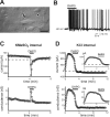
Figure 1.
Figure 1.
Two distinct ionic mechanisms can contribute to mGluR1/5 receptor-mediated increase in striatal interneuron excitability. A, IR-DIC image of a large striatal interneuron directly visualized in a brain slice (arrow); smaller medium spiny cells are also apparent (arrowheads). Scale bar, 20 μm. B, Under whole-cell current clamp, 3,5-DHPG (100 μm), an mGluR1/5-selective agonist, reversibly increased neuronal firing. Resting membrane potential was set to −60 mV by intracellular current injection. Under control conditions, the neuron was silent or fired only sporadically. C, D, Under whole-cell voltage clamp, neurons were held at −60 mV, and hyperpolarizing ramps (Δ−0.05 mV/ms) were applied every 5 s. Representative time series show the effect of 3,5-DHPG on holding current (top) and slope conductance (bottom; from −60 to −80 mV) in a striatal interneuron while recording with either a KMeSO3-based (C) or a KCl-based (D) internal solution. With the KMeSO3-based internal solution, 3,5-DHPG caused a rapidly desensitizing inward current accompanied by an increase in conductance. During recordings with a KCl-based pipette solution, a time-dependent increase in holding current and conductance was observed, which peaked within ∼2 min after whole-cell access; at that point, 3,5-DHPG caused a decrease in outward holding current that was associated with a decrease in conductance. Insets, The first 4 min of the recordings are shown on an expanded time scale.
Figure 2.
Figure 2.
mGluR1/5 receptors activate TrpC-like currents in striatal cholinergic interneurons with pharmacological properties of TrpC3 or TrpC7. A–C, Time series illustrating the effects of histamine (HA; 10 μm) and 3,5-DHPG (50 μm) on holding current at −60 mV (left); I–V relationships of agonist-activated currents were derived by subtracting control ramp currents from those obtained in the presence of agonist (right). A, In striatal interneurons recorded with a KMeSO3-based internal solution, both HA and 3,5-DHPG induced a rapid inward shift in current; the agonist-induced currents did not reverse between −60 and −130 mV. B, When recorded with a CsMeSO3-based internal solution, HA and 3,5-DHPG induced a rapidly desensitizing inward current; examination of agonist-sensitive currents through more depolarized potentials revealed a doubly rectifying I–V profile that reversed at approximately −10 mV. C, In the presence of FFA (500 μm), the 3,5-DHPG-activated current was essentially eliminated. D, Averaged data showing effects of different TrpC channel inhibitors (0 Na+, 100 μm Gd3+, 100 μm La3+, and 500 μm FFA) on mGluR1/5 receptor-activated current; data are expressed as the amplitude ratio of 3,5-DHPG current to the histamine current measured in the same cell (IDHPG:Ihistamine). The combined pharmacological profile is consistent with TrpC3 and TrpC7 channels. *p < 0.0001 by ANOVA.
Figure 3.
Figure 3.
Expression of TrpC3 and TrpC7, but not TrpC6, in striatal cholinergic interneurons. A, B, TrpC3, TrpC6, and TrpC7 transcripts were detected in horizontal sections through the rat brain by in situ hybridization using [33P]-labeled cRNA probes. A, Film autoradiographs show differential expression of these channels throughout the CNS, with prominent expression of TrpC3 and TrpC7 in hippocampus, cerebellum, cortex, and striatum. B, Left, Within the striatum, a punctate distribution reminiscent of cholinergic interneurons (arrows) was evident for TrpC3 and TrpC7, which was particularly obvious by examination of dark-field images of emulsion-dipped sections. Right, At higher magnification, under bright field, heavy accumulations of silver grains can be seen overlying large neurons (arrows), but not on the more numerous and smaller striatal cells. C, Immunohistochemistry for ChAT (left) was combined with nonisotopic in situ hybridization (right) using digoxigenin-labeled cRNA probes for TrpC3 (top) and TrpC7 (bottom); cholinergic (i.e., ChAT immunoreactive) neurons express both TrpC3 and TrpC7. Scale bars: A, 1 cm; B, left, 200 μm; B, right, C, 50 μm.
Figure 4.
Figure 4.
TrpC3 and TrpC7 are activated by both group I mGluRs, mGluR1 and mGluR5. HEK293T cells were cotransfected with either TrpC3 or TrpC7 and mGluR1 or mGluR5. Cells were held at −60 mV, and depolarizing ramps (Δ0.13 mV/ms) were applied at 5 s intervals under control conditions and during exposure to the mGluR1/5 agonist, 3,5-DHPG (50 μm). Ramp I–V curves are presented from the control period and during exposure to the receptor agonist, as indicated, for all combinations of channel and receptor. In all cases, activation of mGluR1 or mGluR5 activated either TrpC3 and TrpC7, as evidenced by the 3,5-DHPG-induced increase in inward and outward current (n ≥ 5 for each combination of receptor and channel).
Figure 5.
Figure 5.
Chloride activates an outwardly rectifying K+ current in striatal interneurons that is inhibited by mGluR1/5 receptors and inhalational anesthetics. Neurons were held at −60 mV, and hyperpolarizing ramps (Δ−0.05 mV/ms) were applied every 5 s while recording with a Cl-based internal solution. A, Subtraction of ramp I–V curves taken immediately after whole-cell access and 100–200 s later shows activation of an outwardly rectifying K+ current. Inset, Time course of the increase in holding current (at −60 mV). B, C, Modulation of the chloride-activated K+ current. Subtracted ramp I–V curves illustrate an outwardly rectifying K+ current inhibited by 50 μm 3,5-DHPG (B) and by 1.9% isoflurane (C) in cells recorded with KCl-based internal solutions. Insets, Time courses depicting the initial increase in holding current and the subsequent inhibition of that run-up current by 3,5-DHPG or isoflurane.
Figure 6.
Figure 6.
Receptor-sensitive cationic and potassium currents in striatal interneuron recorded with gramicidin. A, Representative time series of an interneuron recorded using gramicidin perforated patch; the cell was held at −60 mV, and hyperpolarizing ramps (Δ−0.05 mV/ms) were applied every 5 s while agonists were applied under control conditions and in the presence of La3+ (100 μm). Effects of 3,5-DHPG (50 μm) and histamine (HA; 10 μm) on holding current (top) and slope conductance (bottom; from −60 mV to −80 mV) are illustrated. B, I–V relationships of 3,5-DHPG-sensitive currents derived from two time points (early and late) under control conditions illustrate a biphasic change in conductance occasionally encountered in these recordings. The early I–V curve reflects activation by 3,5-DHPG of a current that does not reverse between −60 and −130 mV, whereas the late I–V curve is consistent with inhibition by 3,5-DHPG of an outwardly rectifying current. C, Subtracted ramp I–V curves of currents evoked by 3,5-DHPG (red) and histamine (blue) in the presence of La3+ reflect only inhibition of the outwardly rectifying K+ current.
Figure 7.
Figure 7.
Differential expression of Slo2.1 (Slick) and Slo2.2 (Slack) in rat striatum. Slo2.1 and Slo2.2 mRNA were detected in horizontal sections through the rat brain by in situ hybridization using [33P]-labeled cRNA probes. A, B, E, F, Film autoradiographs showed differential expression of Slo2.1 (A) and Slo2.2 (E) in the CNS, with substantially more widespread expression of Slo2.2 mRNA; we observed no labeling with sense control probes (B, F). C, D, G, H, Photomicrographs from emulsion-dipped slides under dark-field (C, G) and bright-field (D, H) illumination present expression patterns of Slo2.1 (C, D) and Slo2.2 (G, H) within the striatum. A distributed population of large striatal cells expressed Slo2.1. The arrow in C indicates the cell shown at higher magnification in D. G, H, Slo2.2 mRNA was more widely localized throughout the striatum (G), as expected for expression in medium spiny neurons; note, however, the presence of a small subpopulation of unlabeled medium-sized cells (see H, arrowhead). LV, Lateral ventricle. Scale bars: A, B, E, F, 2 mm; C, G, 100 μm; D, H, 25 μm.
Figure 8.
Figure 8.
Slo2.1 (Slick) is expressed in striatal cholinergic interneurons. A–D, Nonisotopic in situ hybridization using digoxigenin-labeled antisense (A, C) and sense (B, D) cRNA probes (left) for Slo2.1 (A, B) and Slo2.2 (C, D) was combined with immunohistochemistry for ChAT (right) in coronal sections of rat striatum. ChAT-immunopositive striatal cholinergic interneurons labeled with the Slo2.1 probe are indicated (arrows); noncholinergic cortical neurons expressing Slo2.1 are also identified (A, arrowhead). Slo2.2 labeling was not observed in ChAT-immunoreactive striatal interneurons (C; expanded view of the boxed area is provided as an inset). No labeling for Slo2.1 or Slo2.2 was obtained with either of the sense strand control probes. ec, External capsule. Scale bar, 50 μm.
Figure 9.
Figure 9.
Slo2.1 is inhibited by isoflurane and mGluR1/5 receptor activation. HEK293T cells were cotransfected with a GFP-Slick fusion construct and either the mGluR1 or mGluR5 receptor. Cells were held at −60 mV, and depolarizing ramps (Δ0.2 mV/ms) were applied every 5 s under control conditions and during exposure to isoflurane (A) or the mGluR1/5 agonist, 3,5-DHPG (B). Representative time series (left) illustrate inhibition of rSlo2.1 conductance (measured between −80 and −35 mV) by isoflurane and mGluR5 receptor activation. Ramp I–V curves (right) are presented from the control period and during exposure to the drugs, as indicated.

References

    1. Annoura H, Fukunaga A, Uesugi M, Tatsuoka T, Horikawa Y. A novel class of antagonists for metabotropic glutamate receptors, 7-(hydroxyimino)cyclopropa[b]chromen-1a-carboxylates. Bioorg Med Chem Lett. 1996;6:763–766.
    1. Bell MI, Richardson PJ, Lee K. Histamine depolarizes cholinergic interneurones in the rat striatum via a H1-receptor mediated action. Br J Pharmacol. 2000;131:1135–1142. - PMC - PubMed
    1. Bell MI, Richardson PJ, Lee K. Functional and molecular characterization of metabotropic glutamate receptors expressed in rat striatal cholinergic interneurones. J Neurochem. 2002;81:142–149. - PubMed
    1. Bennett BD, Callaway JC, Wilson CJ. Intrinsic membrane properties underlying spontaneous tonic firing in neostriatal cholinergic interneurons. J Neurosci. 2000;20:8493. - PMC - PubMed
    1. Berg AP, Bayliss DA. Striatal cholinergic interneurons express a receptor-insensitive homomeric TASK-3-like background K+ current. J Neurophysiol. 2007;97:1546–1552. - PubMed

Publication types

MeSH terms

LinkOut - more resources