. 2007 Aug 15;27(33):8866-76.
doi: 10.1523/JNEUROSCI.1047-07.2007.

Interaction between telencephalin and ERM family proteins mediates dendritic filopodia formation

Affiliations

Interaction between telencephalin and ERM family proteins mediates dendritic filopodia formation

Yutaka Furutani et al. J Neurosci. .

Abstract

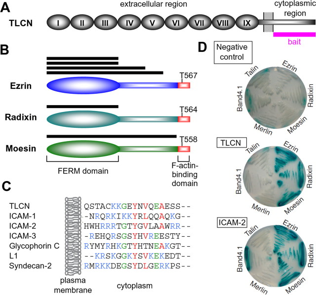
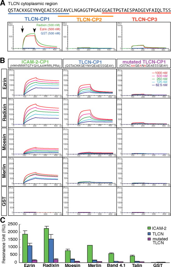
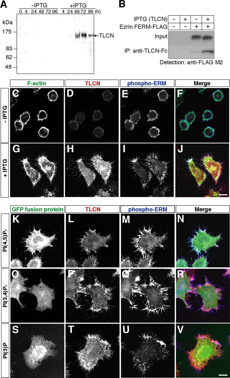
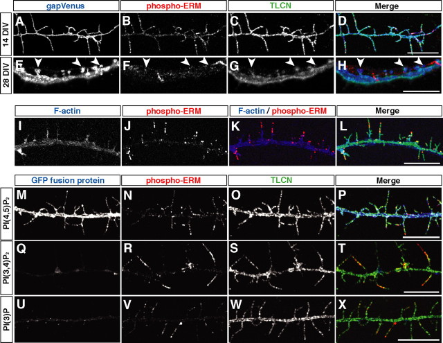
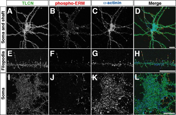
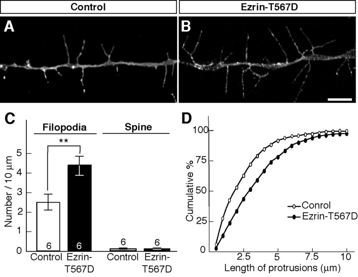
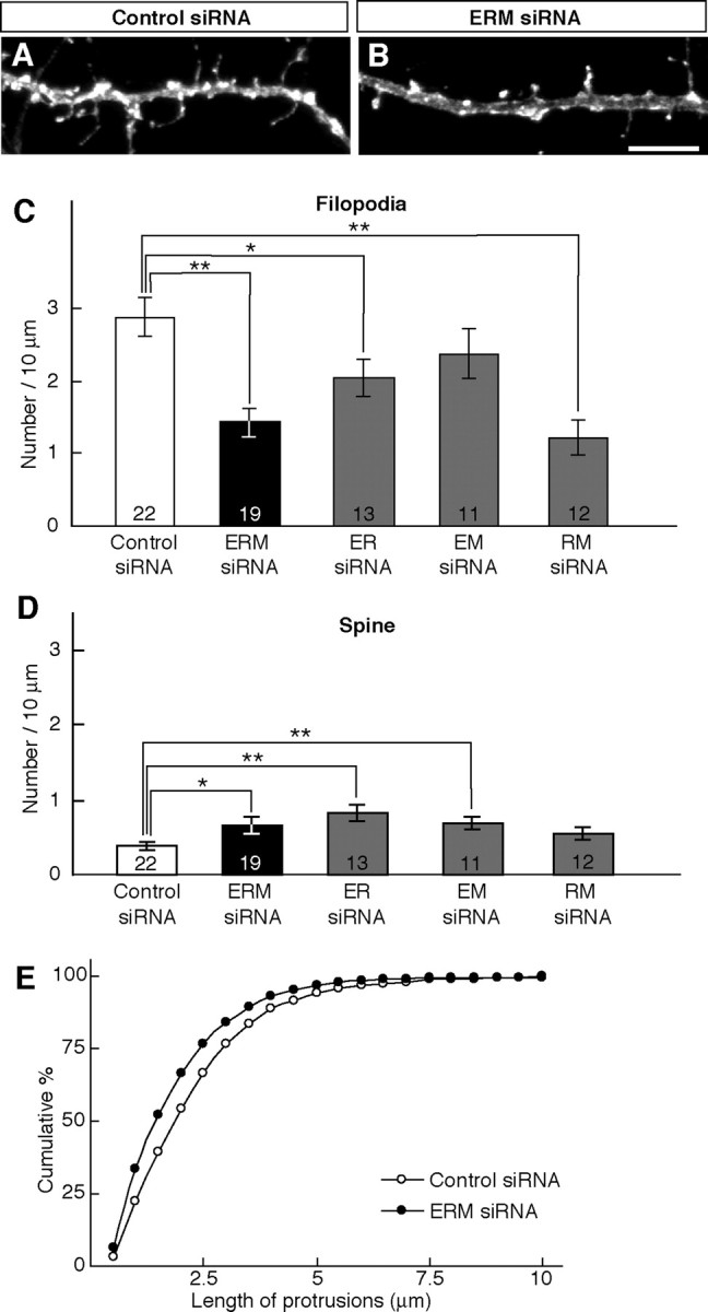
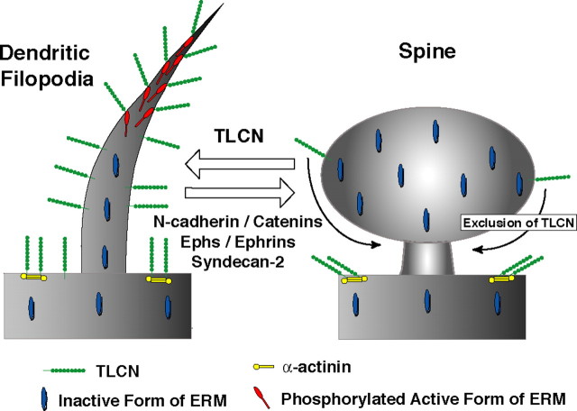
Dendritic filopodia are long, thin, actin-rich, and dynamic protrusions that are thought to play a critical role as a precursor of spines during neural development. We reported previously that a telencephalon-specific cell adhesion molecule, telencephalin (TLCN) [intercellular adhesion molecule-5 (ICAM-5)], is highly expressed in dendritic filopodia, facilitates the filopodia formation, and slows spine maturation. Here we demonstrate that TLCN cytoplasmic region binds ERM (ezrin/radixin/moesin) family proteins that link membrane proteins to actin cytoskeleton. In cultured hippocampal neurons, phosphorylated active forms of ERM proteins are colocalized with TLCN in dendritic filopodia, whereas alpha-actinin, another binding partner of TLCN, is colocalized with TLCN at surface membranes of soma and dendritic shafts. Expression of constitutively active ezrin induces dendritic filopodia formation, whereas small interference RNA-mediated knockdown of ERM proteins decreases filopodia density and accelerates spine maturation. These results indicate the important role of TLCN-ERM interaction in the formation of dendritic filopodia, which leads to subsequent synaptogenesis and establishment of functional neural circuitry in the developing brain.


Figures

Figure 1.
Figure 1.
TLCN cytoplasmic region binds to ERM proteins. A, A schematic diagram depicting the structure of TLCN. Roman numbers indicate nine Ig-like domains in the extracellular region. A whole cytoplasmic region used as a bait for yeast two-hybrid screening is shown as a magenta bar. B, Structures of ezrin, radixin, and moesin. The N-terminal FERM domains and the C-terminal F-actin-binding domains are shown in ovals and red boxes, respectively. T567, T564, and T568 represent phosphorylation sites (Thr residues) near the C terminus. Horizontal black bars indicate positive clones obtained in the two-hybrid screening, all of which contain the N-terminal FERM domains. C, Amino acid sequence alignment of the juxtamembrane region of representative ERM-binding proteins. Hydrophobic residues important for the interaction of ICAM-2 with radixin (Hamada et al., 2003) are shown in red, well conserved Gly and Glu in green, and basic residues in blue. D, Confirmation of the interaction between TLCN and ERM proteins. Yeasts were cotransformed with three expression plasmids: pJGC containing the FERM domain of ezrin, radixin, moesin, merlin, band 4.1 or talin, pEG202 containing TLCN or ICAM-2 cytoplasmic regions or pEG202 (negative control), and a LacZ reporter pSH18–34. Interactions were measured by β-galactosidase activities with 5-bromo-4-chloro-3-indolyl-β-d-galactopyranoside as a substrate.
Figure 2.
Figure 2.
Surface plasmon resonance analysis of binding between TLCN and ERM proteins. A, The amino acid sequence of mouse TLCN cytoplasmic region is shown with three synthetic peptides (TLCN-CP1, TLCN-CP2, and TLCN-CP3; underlines) used for the surface plasmon resonance analysis (Biacore 2000). Biotinylated peptides were immobilized on streptavidin-coated sensor chips, and recombinant GST-fusion proteins containing ezrin and radixin FERM domains (500 nm) were applied. The start and end points of injection of recombinant proteins are indicated by arrow and arrowhead, respectively. The juxtamembrane sequence of TLCN cytoplasmic region specifically binds the FERM domains of ezrin and radixin. B, Specificity and kinetics of binding between TLCN and ERM proteins. Biotinylated ICAM-2-CP1, TLCN-CP1, and mutated TLCN-CP1 peptides were immobilized on streptavidin-coated sensor chips, and serially diluted recombinant GST-fusion proteins containing various FERM domains (1000, 500, 250, 125, and 62.5 nm) were applied. C, Bar graphs summarizing the maximal resonance units for individual interactions between ICAM-2 or TLCN peptides and FERM domains. Error bars indicate SD (n = 3).
Figure 3.
Figure 3.
TLCN induces phospho-ERM-containing filopodia-like protrusions in N2a cells. A, Western blot analysis for the time course of TLCN expression induced by IPTG in TLCN-N2a cells. B, Coimmunoprecipitation of TLCN with FLAG-tagged ezrin FERM domain in TLCN-N2a cells. TLCN-N2a cells were transfected with control or FLAG-tagged ezrin FERM domain-expressing plasmids in the presence or absence of IPTG, lysed, immunoprecipitated with anti-TLCN/Fc antibody, and immunoblotted with anti-FLAG M2 antibody. Input indicates the lysate of cells subjected to immunoprecipitation. C–J, Triple labeling of TLCN/N2a cells in the absence (C–F) or presence (G–J) of IPTG with rhodamine–phalloidin (C, F, G, J; green), anti-TLCN antibody (D, F, H, J; red), and anti-phospho-ERM antibody (E, F, I, J; blue). Merged images are shown in F and J. TLCN expression induces the formation of filopodia-like protrusions containing F-actin and phospho-ERM. K–V, Comparison of localizations of phosphoinositides (K, N, O, R, S, V; green), TLCN (L, N, P, R, T, V; red), and phospho-ERM (M, N, Q, R, U, V; blue) in TLCN/N2a cells in the presence of IPTG. Merged images are shown in N, R, and V. Localizations of phosphoinositides were visualized by transfection of plasmids expressing fluorescent probes, PLCδ1-PH–GFP (K, N), TAPP2-PH–GFP (O, R), and EEA1–3×–FYVE–GFP (S, V), that are specific for PI(4,5)P2, PI(3,4)P2, and PI(3)P, respectively (Balla and Varnai, 2002). Only PI(4,5)P2 is colocalized in TLCN-induced phospho-ERM-containing filopodia-like protrusions. Scale bars, 10 μm.
Figure 4.
Figure 4.
TLCN colocalizes with phospho-ERM in hippocampal neurons. A–H, Triple immunolabeling of gapVenus-expressing cultured hippocampal neurons at 14 DIV (A–D) and 28 DIV (E–H) with anti-GFP (A, D, E, H; blue), anti-phospho-ERM (B, D, F, H; red), and anti-TLCN (C, D, G, H; green) antibodies. Merged images are shown in D and H. I–L, Triple labeling of 14 DIV hippocampal neurons with rhodamine–phalloidin (I, K, L; blue), anti-phospho-ERM antibody (J, K, L; red), and anti-TLCN antibody (L; green). Merged image are shown in K and L. F-actin and phospho-ERM are detected in dendritic filopodia but show distinct localizations; F-actin in proximal and phospho-ERM in distal segments. M–X, Comparison of localizations of phosphoinositides [PI(4,5)P2 in M and P; PI(3,4)P2 in Q and T; PI(3)P in U and X; blue], phospho-ERM (N, P, R, T, V, X; red), and TLCN (O, P, S, T, W, X; green) in hippocampal neurons. Merged images are shown in P, T, and X. Dendritic filopodia contain PI(4,5)P2 together with phospho-ERM and TLCN but not PI(3,4)P2 and PI(3)P. Scale bars, 10 μm.
Figure 5.
Figure 5.
Colocalization of TLCN with phospho-ERM and α-actinin in distinct subcellular domains of hippocampal neurons. A–L, Triple immunofluorescence labeling of hippocampal neurons (14 DIV) with antibodies against TLCN (A, D, E, H, I, L; green), phospho-ERM (B, D, F, H, J, L; red), and α-actinin (C, D, G, H, K, L; blue). E–L, Higher-magnification images of a dendrite (E–H) and a soma (I–L) (indicated as dashed squares in A). Phospho-ERM is preferentially localized to dendritic filopodia (F, H), whereas α-actinin is mostly distributed in soma and dendritic shafts (G, H, K, L). Scale bars: D, 20 μm; H, L, 10 μm.
Figure 6.
Figure 6.
Constitutively active ezrin enhances dendritic filopodia formation. Hippocampal neurons (12 DIV) were injected with control (A) and ezrin–T567D-expressing (B) plasmids, together with gapVenus expressing-plasmid to visualize fine dendritic morphology. The neurons were stained with anti-GFP antibody at 14 DIV. A, B, Representative dendritic morphology of control and ezrin–T567D-expressing neurons. Ezrin–T567D-expressing neurons have more numerous and longer dendritic filopodia than control neurons. Scale bar, 5 μm. C, Densities of filopodia and spines in control (n = 6) and ezrin–T567D-expressing (n = 6) neurons. **p < 0.01, two-tailed Student's t test. Error bars indicate SEM. D, Cumulative frequency plots for dendritic protrusion length in control (open circles) and ezrin–T567D-expressing (filled circles) neurons.
Figure 7.
Figure 7.
ERM knockdown causes malformation of dendritic filopodia. Hippocampal neurons (12 DIV) were injected with 0.75 μg/μl control siRNA-expressing (A), 0.25 μg/μl each of ezrin/radixin/moesin siRNA-expressing (B; ERM siRNA mix), ezrin/radixin/control, ezrin/moesin/control, and radixin/moesin/control siRNA-expressing (ER, EM, and RM siRNA mix) plasmids, together with 0.05 μg/μl gapVenus expression plasmid. The neurons were stained with anti-GFP antibody at 14 DIV. A, B, Representative dendritic morphology of control siRNA- and ERM siRNA mix-expressing neurons. ERM siRNA mix-expressing neurons show the decrease in filopodia number and the acceleration of spine maturation. Scale bar, 5 μm. C, D, Densities of filopodia (C) and spines (D) in control siRNA-expressing (white bars; n = 22), ERM siRNA mix-expressing (black bars; n = 19), ER siRNA mix-expressing (gray bars; n = 13), EM siRNA mix-expressing (gray bars; n = 11), and RM siRNA mix-expressing (gray bars; n = 12) neurons. *p < 0.05 and **p < 0.01, two-tailed Student's t test. Error bars indicate SEM. E, Cumulative frequency plot for dendritic protrusion length in control siRNA-expressing (open circles) and ERM siRNA mix-expressing (filled circles) neurons.
Figure 8.
Figure 8.
A model for the role of TLCN–ERM interaction in dendritic filopodia. In dendritic filopodia, ERM proteins are phosphorylated to become the active form (red) that can bind TLCN (green) in the plasma membrane. In dendritic shafts and spines, inactive ERM proteins (blue) are diffusely present in cytoplasm. Conversely, α-actinin (yellow) constantly binds TLCN in dendritic shafts. The interaction between TLCN and phospho-ERM mediates the formation, elongation, and maintenance of dendritic filopodia and slows spine maturation, whereas N-cadherin/catenins, Ephs/ephrins, and syndecan-2 enhance stabilization of mature synapses.

Similar articles

Cited by

References

    1. Balla T, Varnai P. Visualizing cellular phosphoinositide pools with GFP-fused protein-modules. Sci STKE. 2002;2002:L3. - PubMed
    1. Baumgartner M, Sillman AL, Blackwood EM, Srivastava J, Madson N, Schilling JW, Wright JH, Barber DL. The Nck-interacting kinase phosphorylates ERM proteins for formation of lamellipodium by growth factors. Proc Natl Acad Sci USA. 2006;103:13391–13396. - PMC - PubMed
    1. Benson DL, Yoshihara Y, Mori K. Polarized distribution and cell type-specific localization of telencephalin, an intercellular adhesion molecule. J Neurosci Res. 1998;52:43–53. - PubMed
    1. Bretscher A, Edwards K, Fehon RG. ERM proteins and merlin: integrators at the cell cortex. Nat Rev Mol Cell Biol. 2002;3:586–599. - PubMed
    1. Castelo L, Jay DG. Radixin is involved in lamellipodial stability during nerve growth cone motility. Mol Biol Cell. 1999;10:1511–1520. - PMC - PubMed

Publication types

MeSH terms