. 2007 Aug 15;27(33):8877-84.
doi: 10.1523/JNEUROSCI.2063-07.2007.

Failure to regulate: counterproductive recruitment of top-down prefrontal-subcortical circuitry in major depression

Affiliations

Failure to regulate: counterproductive recruitment of top-down prefrontal-subcortical circuitry in major depression

Tom Johnstone et al. J Neurosci. .

Abstract

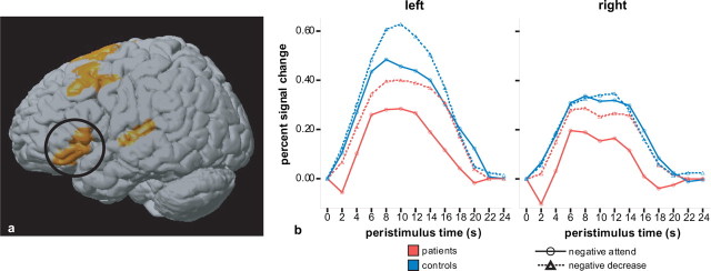
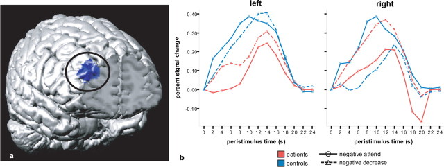
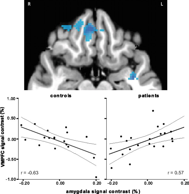
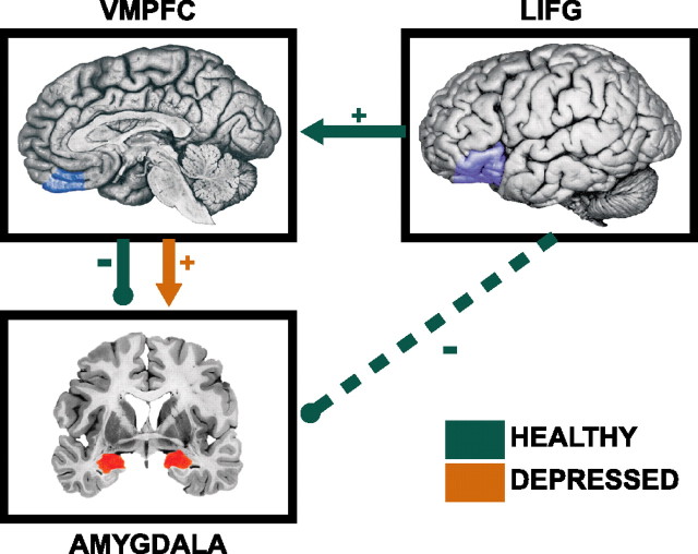
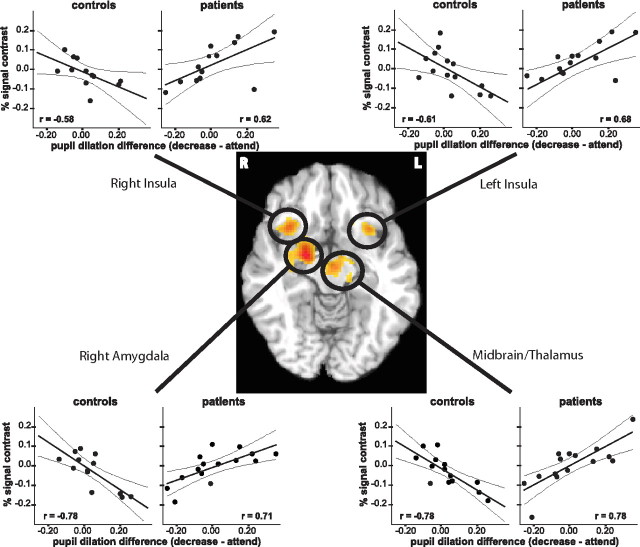
Although depressed mood is a normal occurrence in response to adversity in all individuals, what distinguishes those who are vulnerable to major depressive disorder (MDD) is their inability to effectively regulate negative mood when it arises. Investigating the neural underpinnings of adaptive emotion regulation and the extent to which such processes are compromised in MDD may be helpful in understanding the pathophysiology of depression. We report results from a functional magnetic resonance imaging study demonstrating left-lateralized activation in the prefrontal cortex (PFC) when downregulating negative affect in nondepressed individuals, whereas depressed individuals showed bilateral PFC activation. Furthermore, during an effortful affective reappraisal task, nondepressed individuals showed an inverse relationship between activation in left ventrolateral PFC and the amygdala that is mediated by the ventromedial PFC (VMPFC). No such relationship was found for depressed individuals, who instead show a positive association between VMPFC and amygdala. Pupil dilation data suggest that those depressed patients who expend more effort to reappraise negative stimuli are characterized by accentuated activation in the amygdala, insula, and thalamus, whereas nondepressed individuals exhibit the opposite pattern. These findings indicate that a key feature underlying the pathophysiology of major depression is the counterproductive engagement of right prefrontal cortex and the lack of engagement of left lateral-ventromedial prefrontal circuitry important for the downregulation of amygdala responses to negative stimuli.


Figures

Figure 1.
Figure 1.
Activation in IFG when downregulating emotional responses to negative pictures versus attending to negative pictures. a, Cluster in left IFG shows decrease-attend activation for healthy controls and depressed individuals. Activation in the right IFG is attributable to the depressed group but not the control group having decrease-attend activation in this region. Talairach coordinates: x ± 47, y = 24, z = 0, BA 45/47. b, Mean peristimulus plots of estimated BOLD responses to negative pictures in the attend condition (solid line) and the decrease condition (dashed line). The control group is shown in blue, and the depressed group is shown in red. The activation was left-lateralized in controls but bilateral in the depressed group (group × regulation × hemisphere interaction for cluster mean, F (1,37) = 4.26; p = 0.046).
Figure 2.
Figure 2.
Cluster in the right MFG showing a group by regulation interaction. a, Cluster location Talairach coordinates: x = 32, y = 46, z = 25, BA 10. b, Peristimulus time plots of estimated BOLD responses as a function of group and regulation condition. In the right MFG, controls showed a decrease in activation when decreasing responses to negative pictures (relative to attending), whereas the depressed individuals showed an increase in activation when decreasing their responses (group × condition × hemisphere interaction for cluster mean: F (1,37) = 8.01; p = 0.007).
Figure 3.
Figure 3.
Cluster in VMPFC showing significantly different decrease-attend correlation with amygdala for controls versus the depressed group. VMPFC activation correlated negatively with amygdala in controls (r = −0.63; p = 0.005) but correlated positively for the depressed group (r = 0.57; p = 0.007; Talairach coordinates, x = 7, y = 39, z = −11, BA 11/32). R, Right; L, left.
Figure 4.
Figure 4.
Schematic depicting brain regions that showed a significantly different association with amygdala in the control versus depressed individuals. In healthy controls, there was a negative correlation between the decrease-attend contrast in VMPFC and amygdala (r = −0.63; p = 0.005), such that nondepressed individuals who showed the greatest VMPFC activation when downregulating their emotional responses, relative to attending to the pictures, also showed the lowest amygdala activation. The VMPFC was found to be a significant mediator of the association between left IFG (LIFG) and amygdala in controls (Z = −1.86; p = 0.03, one-tailed). In the depressed group, there was a positive correlation in activation between the VMPFC and amygdala (r = 0.57; p = 0.007) when decreasing their emotional response relative to the attend condition, with no mediational effect of VMPFC (Z > 0).
Figure 5.
Figure 5.
Regions showing a significant group difference in the correlation between pupil dilation (decrease-attend) and brain activation (decrease-attend) while reappraising negative pictures. Across all clusters, the same pattern was evident, with a negative correlation in controls and a positive correlation in the depressed group. R, Right; L, left.

Similar articles

Cited by

References

    1. Amaral DG, Price JL. Amygdalo-cortical projections in the monkey (Macaca fascicularis) J Comp Neurol. 1984;230:465–496. - PubMed
    1. Barbas H. Anatomic basis of cognitive-emotional interactions in the primate prefrontal cortex. Neurosci Biobehav Rev. 1995;19:499–510. - PubMed
    1. Beauregard M, Paquette V, Levesque J. Dysfunction in the neural circuitry of emotional self-regulation in major depressive disorder. NeuroReport. 2006;17:843–846. - PubMed
    1. Bernick N, Overlander M. Effect of verbalization and two different modes of experiencing on pupil size. Percept Psychophys. 1968;3:327–330.
    1. Brody AL, Saxena S, Mandelkern MA, Fairbanks LA, Ho ML, Baxter LR. Brain metabolic changes associated with symptom factor improvement in major depressive disorder. Biol Psychiatry. 2001;50:171–178. - PubMed

MeSH terms

LinkOut - more resources