. 2007 Aug 15;27(33):8927-39.
doi: 10.1523/JNEUROSCI.2322-07.2007.

Protein kinase C regulates local synthesis and secretion of a neuropeptide required for activity-dependent long-term synaptic plasticity

Affiliations

Protein kinase C regulates local synthesis and secretion of a neuropeptide required for activity-dependent long-term synaptic plasticity

Jiang-Yuan Hu et al. J Neurosci. .

Abstract

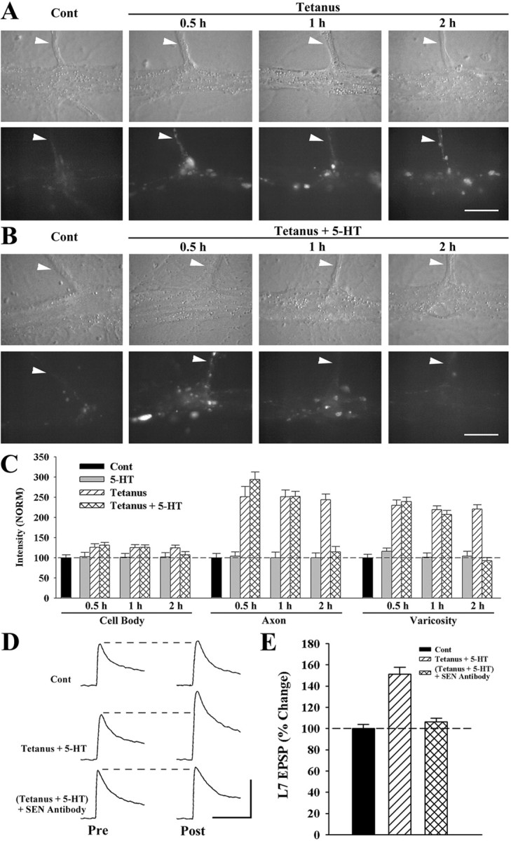
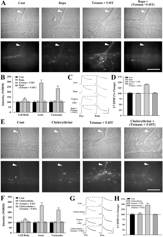
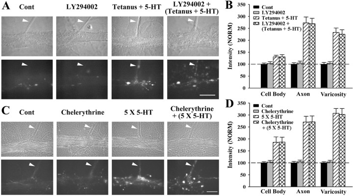
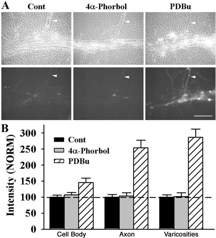
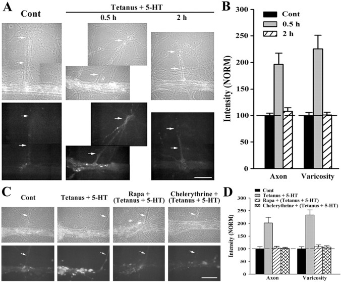
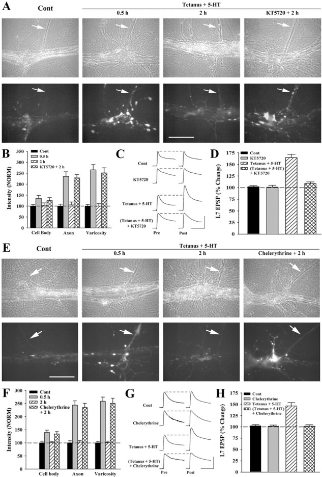
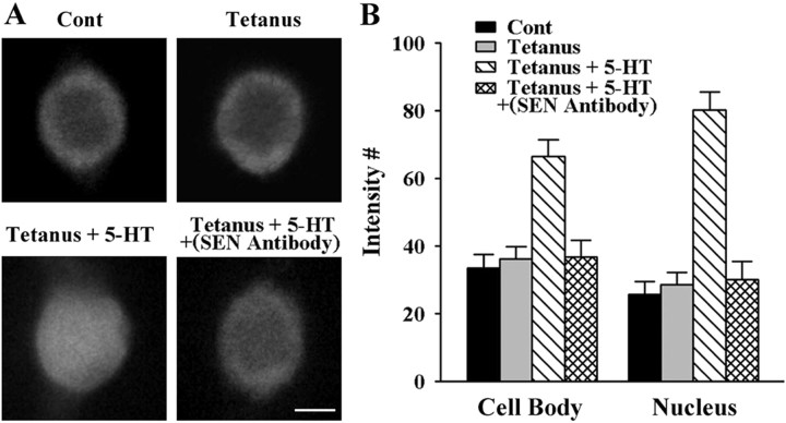
Long-term facilitation (LTF) of sensory neuron synapses in Aplysia is produced by either nonassociative or associative stimuli. Nonassociative LTF can be produced by five spaced applications of serotonin (5-HT) and requires a phosphoinosotide 3-kinase (PI3K)-dependent and rapamycin-sensitive increase in the local synthesis of the sensory neuron neuropeptide sensorin and a protein kinase A (PKA)-dependent increase in the secretion of the newly synthesized sensorin. We report here that associative LTF produced by a single pairing of a brief tetanus with one application of 5-HT requires a rapid protein kinase C (PKC)-dependent and rapamycin-sensitive increase in local sensorin synthesis. This rapid increase in sensorin synthesis does not require PI3K activity or the presence of the sensory neuron cell body but does require the presence of the motor neuron. The secretion of newly synthesized sensorin by 2 h after stimulation requires both PKA and PKC activities to produce associative LTF because incubation with exogenous anti-sensorin antibody or the kinase inhibitors after tetanus plus 5-HT blocked LTF. The secreted sensorin leads to phosphorylation and translocation of p42/44 mitogen-activated protein kinase (MAPK) into the nuclei of the sensory neurons. Thus, different stimuli activating different signaling pathways converge by regulating the synthesis and release of a neuropeptide to produce long-term synaptic plasticity.


Figures

Figure 1.
Figure 1.
Tetanus plus 5-HT increased both sensorin expression and release required for associative LTF. A, B, Nomarski contrast (top) and sensorin immunofluorescent images (bottom) of cocultures 0.5 h after control (Cont) (mock application) or at 0.5, 1, and 2 h after tetanus (2 s at 20 Hz; A) or after tetanus plus 5-HT (2 s at 20 Hz plus 5 min application; B). Staining for sensorin, confined primarily to granules in the sensory neuron axons (arrowheads), regenerated processes or varicosities contacting L7, increased at 0.5 h and 1 h after either stimuli, but decreased to control levels at 2 h only after tetanus plus 5-HT. The axons and processes of L7 are unstained for sensorin. Scale bar, 30 μm. C, Summary of sensorin immunostaining in cell body, axon, and varicosities of sensory neurons after control (Cont) and at various times after 5-HT, tetanus, or tetanus plus 5-HT. Staining in each compartment was normalized to the average staining for that compartment after control (dashed line at 100%). ANOVA indicated a significant effect of treatment (df = 18, 162; F = 48.522; p < 0.001). 5-HT alone produced no significant change in sensorin staining, but tetanus significantly increased staining in axons and varicosities at 0.5 h (F = 5.34 and F = 8.027; p < 0.01), at 1 h (F = 7.004 and F = 8.851; p < 0.01) and at 2 h (F = 6.321 and F = 9.067; p < 0.01). Tetanus plus 5-HT significantly increased staining in axons and varicosities at 0.5 h (F = 12.976 and F = 10.72; p < 0.01) and at 1 h (F = 8.39 and F = 7.185; p < 0.01). Sensorin staining was no longer significantly different from control or 5-HT at 2 h after tetanus plus 5-HT. Sensorin staining in the sensory neuron cell body increased modestly after tetanus or tetanus plus 5-HT but was not significantly different from control or 5-HT. D, E, Sensorin released after tetanus plus 5-HT is required for associative LTF. EPSPs were recorded before (Pre) and 24 h after (Post) control, tetanus plus 5-HT with control Ab applied between 0.5 h and 2 h, or tetanus plus 5-HT with anti-sensorin Ab applied between 0.5 and 2 h (D). The height of the dashed line is the amplitude of each EPSP before treatment. Calibration: 20 mV, 25 ms. Summary of the changes in EPSPs 24 h after treatment (E). ANOVA indicated a significant effect of treatment (df = 2, 30; F = 32.955; p < 0.001). Tetanus plus 5-HT with control Ab produced a significant increase in EPSP amplitude compared with control and with tetanus plus 5-HT with anti-sensorin Ab (F = 25.503 and F = 23.689; p < 0.01). Tetanus plus 5-HT plus anti-sensorin Ab did not significantly affect EPSP amplitude compared with control. The dashed line represents the height of the bar when there was no change in EPSP amplitude.
Figure 2.
Figure 2.
Rapamycin and PKC inhibitor blocked the rapid increase in sensorin and associative LTF produced by tetanus plus 5-HT. A, Nomarski contrast (top) and immunofluorescent images of sensorin staining (bottom) after control (Cont), after 45 min incubation with rapamycin (Rapa), 0.5 h after tetanus plus 5-HT, and 0.5 h after tetanus plus 5-HT with rapamycin. Rapamycin blocked the increase in sensorin staining in both axons (arrowheads) and varicosities produced by tetanus plus 5-HT. Scale bar, 30 μm. B, Summary of sensorin immunostaining after treatments. Staining in each compartment was normalized to the average staining after control (dashed line at 100%). ANOVA indicated a significant effect of treatment (df = 6, 44; F = 10.095; p < 0.001). Compared with control, tetanus plus 5-HT increased significantly sensorin staining in axons and varicosities (F = 22.131 and F = 13.874; p < 0.01). In the presence of rapamycin, tetanus plus 5-HT failed to significantly change sensorin staining compared with controls, and change in staining levels were significantly lower than the change in sensorin staining produced by tetanus plus 5-HT in the absence of drug in the axons and varicosities (F = 21.73 and F = 13.311; p < 0.01). Sensorin staining in the cell body increased modestly after tetanus plus 5-HT but was not significantly different from control or the other treatments. C, D, Rapamycin blocked associative LTF. EPSPs were recorded before (Pre) and 24 h after (Post) treatments (C). Calibration: 20 mV, 25 ms. Summary of the changes in EPSP amplitudes 24 h after treatment (D). ANOVA indicated a significant effect of treatment (df = 3, 20; F = 35.813; p < 0.001). Tetanus plus 5-HT evoked a significant increase in EPSP compared with control or tetanus plus 5-HT with rapamycin (F = 22.682 and F = 25.448; p < 0.01). Tetanus plus 5-HT with rapamycin did not significantly affect EPSP amplitudes compared with control or rapamycin alone. E, Nomarski contrast (top) and immunofluorescent images of sensorin staining (bottom) immediately after control (Cont), after 45 min incubation with chelerythrine, 0.5 h after tetanus plus 5-HT, and 0.5 h after tetanus plus 5-HT with chelerythrine. Chelerythrine blocked the increase in sensorin staining in both axons (arrowheads) and varicosities produced by tetanus plus 5-HT. Scale bar, 30 μm. F, Summary of sensorin immunostaining after treatments. Staining in each compartment was normalized to the average staining after control (dashed line at 100%). ANOVA indicated a significant effect of treatment (df = 6, 44; F = 31.174; p < 0.001). Compared with control, tetanus plus 5-HT produced significant changes in sensorin staining in axons and varicosities (F = 19.454 and F = 17.267; p < 0.01). In the presence of chelerythrine, tetanus plus 5-HT failed to change significantly sensorin staining, and staining levels were significantly lower than the changes in sensorin staining produced by tetanus plus 5-HT without drug both in the axons and varicosities (F = 21.999 and F = 19.013; p < 0.01). G, H, Chelerythrine blocked associative LTF. EPSPs were recorded before (Pre) and 24 h after (Post) treatments (G). Calibration: 20 mV, 25 ms. Summary of the changes in EPSP amplitudes 24 h after treatments (H). ANOVA indicated a significant effect of treatment (df = 3, 23; F = 15.387; p < 0.001). Tetanus plus 5-HT evoked a significant increase in EPSP amplitude compared with control and with tetanus plus 5-HT with chelerythrine (F = 9.62 and F = 12.166; p < 0.01). Tetanus plus 5-HT with chelerythrine did not significantly affect EPSP amplitudes compared with control or chelerythrine alone.
Figure 3.
Figure 3.
Signaling pathway required for regulating rapid sensorin synthesis differs for associative LTF and nonassociative LTF. A, B, PI3K activity is not required for rapid sensorin synthesis after tetanus plus 5-HT. Nomarski contrast (top) and immunofluorescent images of sensorin staining (bottom) after control (Cont), after 45 min incubation with PI3K inhibitor (LY294002), 0.5 h after tetanus plus 5-HT, and 0.5 h after tetanus plus 5-HT with LY294002. The inhibitor failed to block the increase in sensorin staining in both axons (arrowheads) and varicosities produced by tetanus plus 5-HT. Scale bar, 30 μm. B, Summary of sensorin immunostaining after treatments. Staining in each compartment was normalized to the average staining after control (dashed line at 100%). ANOVA indicated a significant effect of treatment (df = 6, 44; F = 28.486; p < 0.001). Compared with control, tetanus plus 5-HT significantly increased sensorin staining in axons and varicosities (F = 14.78 and F = 13.332; p < 0.01). In the presence of LY294002, tetanus plus 5-HT also significantly increased sensorin staining in the axons and varicosities (F = 15.329 and F = 12.833; p < 0.01). Staining after tetanus plus 5-HT with drug was not significantly different from the staining detected after tetanus plus 5-HT without drug. Sensorin staining in the cell body increased modestly after tetanus plus 5-HT but was not significantly different from control or the other treatments. C, D, PKC activity is not required for the increase in sensorin after five applications of 5-HT. Nomarski contrast (top) and immunofluorescent images of sensorin staining (bottom) after control (Cont), after 2 h incubation with chelerythrine, immediately after five applications of 5-HT, and immediately after five applications of 5-HT plus chelerythrine. The inhibitor failed to block the increase in sensorin staining in both axons (arrowheads) and varicosities produced by five applications of 5-HT. Scale bar, 30 μm. B, Summary of sensorin immunostaining after treatments. Staining in each compartment was normalized to the average staining after control (dashed line at 100%). ANOVA indicated a significant effect of treatment (df = 6, 44; F = 41.679; p < 0.001). Compared with control, five applications of 5-HT significantly increased sensorin staining in cell bodies (F = 4.272; p < 0.05) and axons and varicosities (F = 13.421 and F = 23.232; p < 0.01). In the presence of chelerythrine, tetanus plus 5-HT also significantly increased sensorin staining in the cell bodies (F = 4.343; p < 0.05) and axons and varicosities (F = 13.602 and F = 22.281; p < 0.01). Staining after five applications of 5-HT with drug was not significantly different from the staining after five applications of 5-HT without drug.
Figure 4.
Figure 4.
PDBu rapidly increased sensorin in sensory neurons. A, Phase contrast (top) and immunofluorescent images of sensorin staining (bottom) in sensory neuron axons (arrowheads) and varicosities immediately after control treatment (Cont), 0.5 h after 5 min application of inactive phorbol (4α-phorbol), and 0.5 h after 5 min application of PDBu. Sensorin staining increased in the axon and varicosities only after treatment with PDBu. Scale bar, 50 μm. B, Summary of sensorin immunostaining after treatments. Staining in each compartment was normalized to the average staining after control (dashed line at 100%). ANOVA indicated a significant effect of treatment (df = 4, 34; F = 51.524; p < 0.001). PDBu significantly increased sensorin staining compared with control or 4α-phorbol in the sensory neuron cell bodies (F = 5.76, p < 0.03; or F = 4.195, p < 0.05), axons (F = 23.379 or F = 22.376; p < 0.01), and varicosities (F = 26.766 or F = 25.337; p < 0.01). Treatment with 4α-phorbol did not significantly affect sensorin staining compared with control.
Figure 5.
Figure 5.
Rapamycin and PKC inhibitor blocked the increase in sensorin produced by tetanus plus 5-HT in the absence of the sensory neuron cell body. A, B, Without sensory neuron cell bodies, tetanus plus 5-HT rapidly increased sensorin at 0.5 h that recovered to control levels by 2 h. Phase contrast (A, top) and immunofluorescent images of sensorin staining (A, bottom) in sensory neuron axons (bottom arrows in each image) and varicosities after removal of each sensory neuron cell body (former position indicated by the top arrow in each image). After recovery, cultures were treated either with tetanus plus 5-HT or control (Cont). Sensorin staining increased at 0.5 h after tetanus plus 5-HT and returned to control levels by 2 h after stimuli. Scale bar, 50 μm. Summary of sensorin staining in axons and varicosities of sensory neurons without cell bodies after control and 0.5 and 2 h after tetanus plus 5-HT (B). Staining in each compartment was normalized to the average staining after control (dashed line at 100%). ANOVA indicated a significant effect of treatment (df = 2, 17; F = 6.83; p < 0.007). Tetanus plus 5-HT significantly increased sensorin staining at 0.5 h compared with control in both the axons and varicosities (F = 11.86 and F = 14.599; p < 0.01). Sensorin staining in both axons and varicosities 2 h after tetanus plus 5-HT was not significantly different from control and was significantly lower than the staining at 0.5 h after tetanus plus 5-HT in both the axons and varicosities (F = 10.767 and F = 15.53; p < 0.01). C, D, Rapamycin (Rapa) and PKC inhibitor blocked the increase in sensorin staining at 0.5 h after tetanus plus 5-HT. Phase contrast (C, top) and immunofluorescent images of sensorin staining (C, bottom) of cocultures without sensory neuron cell bodies after control application (cont), 0.5 h after tetanus plus 5-HT, 0.5 h after tetanus plus 5-HT with rapamycin, or 0.5 h after tetanus plus 5-HT with chelerythrine. Sensorin staining in axons and varicosities increased only after tetanus plus 5-HT without drug. Scale bar, 50 μm. Summary of sensorin staining in axons and varicosities of sensory neurons without cell bodies after control and 0.5 h after tetanus plus 5-HT with or without drug (D). Staining in each compartment was normalized to the average staining after control (dashed line at 100%). ANOVA indicated a significant effect of treatment (df = 3, 22; F = 5.794; p < 0.005). Tetanus plus 5-HT significantly increased sensorin staining at 0.5 h compared with control in both the axons and varicosities (F = 9.803 and F = 19.429; p < 0.01). Tetanus plus 5-HT with either drug did not significantly affect sensorin staining in the axons or varicosities. Tetanus plus 5-HT without drug increase sensorin staining compared with tetanus plus 5-HT with rapamycin in both axons and varicosities (F = 9.296 and F = 17.253; p < 0.01) and compared with tetanus plus 5-HT with chelerythrine in both axons and varicosities (F = 10.324 and F = 19.019; p < 0.01).
Figure 6.
Figure 6.
Both PKA and PKC activities are required for the release of sensorin after tetanus plus 5-HT and associative LTF. A, B, PKA inhibitor blocked the decline in sensorin staining 2 h after tetanus plus 5-HT. Phase contrast (A, top) and immunofluorescent images of sensorin staining (A, bottom) after control (Cont), 0.5 h after tetanus plus 5-HT, 2 h after tetanus plus 5-HT, and 2 h after tetanus plus 5-HT with KT5720 present between 0.5 and 2 h. The rapid increase in sensorin staining in axons (arrows) and varicosities at 0.5 h does not decline at 2 h with the PKA inhibitor. Scale bar, 50 μm. Summary of the changes in sensorin staining after treatments (B). Staining in each compartment was normalized to the average staining after control (dashed line at 100%). ANOVA indicated a significant effect of treatment (df = 6, 40; F = 37.858; p < 0.001). Compared with control, tetanus plus 5-HT at 0.5 h produced a significant increase in sensorin staining in both the axons and varicosities (F = 13.208 and F = 13.86; p < 0.01). Without PKA inhibitor, sensorin staining in all compartments at 2 h after tetanus plus 5-HT was not significantly different from control. Sensorin staining at 2 h after tetanus plus 5-HT with KT5720 was not significantly different from the staining at 0.5 h after tetanus plus 5-HT, remained significantly higher than control in both the axons and varicosities (F = 11.941 and F = 11.61; p < 0.01), and was significantly higher than the staining 2 h after tetanus plus 5-HT without drug both in the axons and varicosities (F = 11.002 and F = 11.16; p < 0.01). C, D, PKA inhibition between 0.5 and 2 h after tetanus plus 5-HT blocked associative LTF. EPSPs were recorded before (Pre) and 24 h after (post) control, 90 min of KT5720, tetanus plus 5-HT, and tetanus plus 5-HT with KT5720 added between 0.5 and 2 h after paired stimuli (C). Calibration: 20 mV, 25 ms. Summary of the changes in EPSP amplitudes 24 h after treatment (D). ANOVA indicated a significant effect of treatment (df = 3, 23; F = 145.627; p < 0.001). Change after tetanus plus 5-HT with KT5720 was not significantly different from control or treatment with KT5720 alone but was significantly lower than the increase produced tetanus plus 5-HT without drug (F = 26.509; p < 0.01). Tetanus plus 5-HT significantly increased EPSP amplitude compared with control (F = 29.108; p < 0.01). E, F, PKC inhibitor blocked the decline in sensorin staining 2 h after tetanus plus 5-HT. Phase contrast (E, top) and immunofluorescent images of sensorin staining (E, bottom) after control, 0.5 h after tetanus plus 5-HT, 2 h after tetanus plus 5-HT, and 2 h after tetanus plus 5-HT with chelerythrine present between 0.5 and 2 h. The rapid increase in sensorin staining in axons (arrows) and varicosities at 0.5 h does not decline at 2 h with the PKC inhibitor. Scale bar, 50 μm. Summary of the changes in sensorin staining after treatments (F). Staining in each compartment was normalized to the average staining after control (dashed line at 100%). ANOVA indicated a significant effect of treatment (df = 6, 48; F = 42.206; p < 0.001). Compared with control, tetanus plus 5-HT at 0.5 h produced a significant increase in sensorin staining in the sensory neuron cell bodies (F = 3.951; p < 0.05) and axons and varicosities (F = 18.05 and F = 18.986; p < 0.01). Without PKC inhibitor, sensorin staining in all compartments at 2 h after tetanus plus 5-HT was not significantly different from control. Sensorin staining at 2 h after tetanus plus 5-HT with chelerythrine was not significantly different from the staining at 0.5 h after tetanus plus 5-HT, remained significantly higher than control both in the axons and varicosities (F = 16.071 and F = 18.052; p < 0.01), and was significantly higher than the staining 2 h after tetanus plus 5-HT without drug in both the axons and varicosities (F = 17.563 and F = 19.396; p < 0.01). G, H, PKC inhibition between 0.5 and 2 h after tetanus plus 5-HT blocked associative LTF. EPSPs were recorded before (Pre) and 24 h after (Post) control, 90 min of chelerythrine, tetanus plus 5-HT, and tetanus plus 5-HT with chelerythrine added between 0.5 and 2 h after paired stimuli (G). Calibration: 20 mV, 25 ms. Summary of the changes in EPSP amplitudes 24 h after treatment (H). An ANOVA indicated a significant effect of treatment (df = 3, 22; F = 125.062; p < 0.001). Change in EPSP amplitude after tetanus plus 5-HT with chelerythrine was not significantly different from control or treatment with chelerythrine alone but was significantly lower than the increase produced by tetanus plus 5-HT without drug (F = 18.471; p < 0.01). Tetanus plus 5-HT significantly increased EPSP amplitude compared with control (F = 15.48; p < 0.01).
Figure 7.
Figure 7.
Activation and translocation of p42/44 MAPK in the cell bodies of sensory neurons 1 h after tetanus plus 5-HT is blocked by anti-sensorin Ab. A, Immunofluorescent images of phospho MAPK staining in the cell body of sensory neurons 1 h after control (Cont), tetanus, tetanus plus 5-HT, and tetanus plus 5-HT with incubation for 1 h with anti-sensorin (SEN) Ab. Note the increase in overall staining in the cell body and staining in the nucleus only after tetanus plus 5-HT. Scale bar, 25 μm. B, Summary of phospho-MAPK immunostaining in the whole cell body and nucleus after treatments. ANOVA indicated a significant effect of treatment (df = 3, 38; F = 35.255; p < 0.001). Tetanus plus 5-HT significantly increased staining in both the cell body and nucleus compared with control (F = 8.992 and F = 22.317; p < 0.01), tetanus alone (F = 7.234 and F = 18.89; p < 0.01), and tetanus plus 5-HT with anti-sensorin Ab (F = 7.748 and F = 19.843; p < 0.01). There was no significant difference in staining in the cell body or nucleus after tetanus plus 5-HT with anti-sensorin Ab compared with control or tetanus alone.

Similar articles

Cited by

References

    1. Abrams TW, Karl KA, Kandel ER. Biochemical studies of stimulus convergence during classical conditioning in Aplysia: dual regulation of adenylate cyclase by calcium/calmodulin and transmitter. J Neurosci. 1991;11:2655–2665. - PMC - PubMed
    1. Abrams TW, Yovell Y, Onyike CU, Cohen JE, Jarrard HE. Analysis of sequence-dependent interactions between transient calcium and transmitter stimuli in activating adenylyl cyclase in Aplysia: possible contribution to CS-US sequence requirement during conditioning. Learn Mem. 1998;4:496–509. - PubMed
    1. Alberini CM, Ghirardi M, Metz R, Kandel ER. C/EBP is an immediated-early gene required for the consolidation of long-term facilitation in Aplysia. Cell. 1994;76:1099–1114. - PubMed
    1. Angers S, Fioravante D, Chin J, Cleary LJ, Bean AJ, Byrne JH. Serotonin stimulates phosphorylation of Aplysia synapsin and alters its subcellular distribution. J Neurosci. 2002;22:5412–5422. - PMC - PubMed
    1. Bacskai BJ, Hochner B, Mahaut-Smith M, Adams SR, Kaang BK, Kandel ER, Tsien RY. Spatially resolved dynamics of cAMP and protein kinase A subunits in Aplysia sensory neurons. Science. 1993;260:222–226. - PubMed

Publication types

MeSH terms

LinkOut - more resources