. 2007 Aug 15;27(33):8940-51.
doi: 10.1523/JNEUROSCI.2085-07.2007.

Dominant-negative inhibition of M-like potassium conductances in hair cells of the mouse inner ear

Affiliations

Dominant-negative inhibition of M-like potassium conductances in hair cells of the mouse inner ear

Jeffrey R Holt et al. J Neurosci. .

Abstract

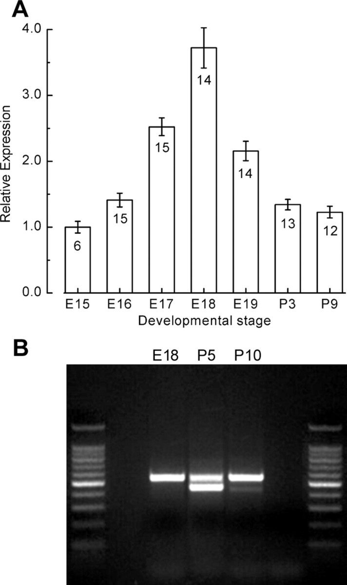
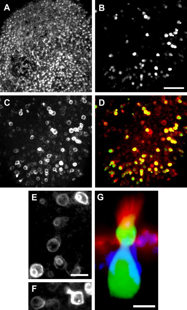
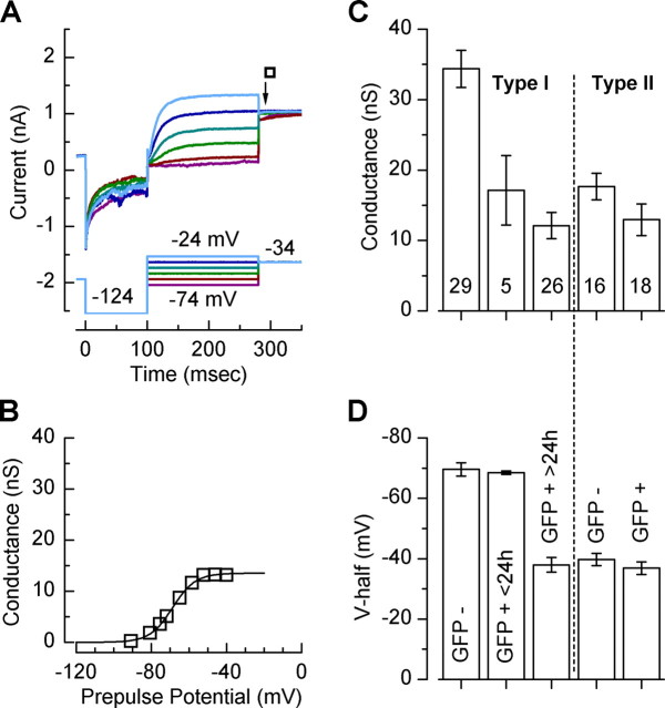
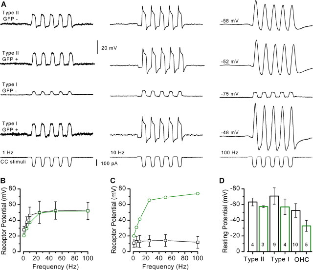
Sensory hair cells of the inner ear express multiple physiologically defined conductances, including mechanotransduction, Ca(2+), Na(+), and several distinct K(+) conductances, all of which are critical for normal hearing and balance function. Yet, the molecular underpinnings and their specific contributions to sensory signaling in the inner ear remain obscure. We sought to identify hair-cell conductances mediated by KCNQ4, which, when mutated, causes the dominant progressive hearing loss DFNA2. We used the dominant-negative pore mutation G285S and packaged the coding sequence of KCNQ4 into adenoviral vectors. We transfected auditory and vestibular hair cells of organotypic cultures generated from the postnatal mouse inner ear. Cochlear outer hair cells and vestibular type I cells that expressed the transfection marker, green fluorescent protein, and the dominant-negative KCNQ4 construct lacked the M-like conductances that typify nontransfected control hair cells. As such, we conclude that the M-like conductances in mouse auditory and vestibular hair cells can include KCNQ4 subunits and may also include KCNQ4 coassembly partners. To examine the function of M-like conductances in hair cells, we recorded from cells transfected with mutant KCNQ4 and injected transduction current waveforms in current-clamp mode. Because the M-like conductances were active at rest, they contributed to the very low potassium-selective input resistance, which in turn hyperpolarized the resting potential and significantly attenuated the amplitude of the receptor potential. Modulation of M-like conductances may allow hair cells the ability to control the amplitude of their response to sensory stimuli.


Figures

Figure 1.
Figure 1.
Expression of KCNQ4 mRNA in the mouse utricle. A, Quantitative real-time RT-PCR was used to examine the expression pattern of KCNQ4 in the developing mouse utricle. A standard curve was generated using tenfold dilution series of plasmid DNA that carried the coding sequence for mKCNQ4 was used to confirm primer efficiency. The relative expression levels are plotted as a function of developmental stage. The number of samples and SEs are shown for each stage. B, Image of an agarose gel electrophoresis showing the amplification products of an RT-PCR using primers that span exons 8 through 12 of mKCNQ4 and template harvested from the developmental stages indicated. A 1 kb ladder was run in lanes 1 and 5. Bands of 528 and 423 base pairs were amplified at P5 and P10.
Figure 2.
Figure 2.
Generation and validation of adenoviral expression of mutant KCNQ4. A, Vector maps of the expression cassette introduced into the three adenoviral vectors used for this study. Promoter sequences are shown in blue, GFP in green, and the coding sequence of KCNQ4 in yellow. The name assigned to each vector is shown at the left. B, A family of currents recorded from HEK293 cells stably transfected with hKNCQ4 (Sogaard et al., 2001). The voltage protocol is shown below. C, The stably transfected cells were exposed to 1 × 10⁵ viral particles/ml of A2-mKCNQ4-G285S for 4 h. Twenty-four hours later, GFP-positive cells were identified. Shown is a representative family of currents evoked by the protocol shown below. D, DIC and fluorescence images from an A2-mKCNQ4-G285S-transfected HEK293 cell. E, Activation curves derived from the data shown in B and C. The wild-type hKNCQ4 (circles) and mutant mKCNQ4-G285S (diamonds) were fit with Boltzmann curves that had the following parameters, respectively: Gmax, 25.8 nS; V(1/2), −34 mV; s, 18.6 mV; Gmax, 3.2 nS; V(1/2), −43 mV; s, 11.9 mV.
Figure 3.
Figure 3.
Confocal images of a P8 mouse utricle explant 48 h after transfection with 8.6 × 10⁸ particles/ml of Ad1-VgRXR and 1.4 × 10¹⁰ particles/ml of Ad1-ecd-hKCNQ4-G285S. A, Top-down view of the whole-mount utricle with the actin-rich hair bundles illuminated with phalloidin-Alexa546. A z-series that consisted of 20 focal planes was collapsed to generate images A–F. B, The GFP signal from the same field as shown in A. C, The same field as shown in A and B stained with anti-KCNQ4 and an Alexa633 secondary antibody. D, Merge of B and C with GFP shown in green and KCNQ4 shown in red. E, Close-up view of several KCNQ4-postive cells. The cells in the top right and lower left were also positive for GFP. Note the ring within a ring pattern of fluorescence that characterized both the transfected type I cells and the nontransfected type I cells (bottom center). F, Close-up view of a GFP-positive type I cell (right) that reveals the restricted neck region. G, High-magnification cross-sectional view of a GFP-positive type I cell with GFP in green, phalloidin in red, and KNCQ4 in blue. Scale bars: (in B) A–D, 50 μm; E, F, 10 μm. G, 5 μm.
Figure 4.
Figure 4.
Representative control potassium currents recorded from nontransfected vestibular hair cells. A, A family of control currents recorded from nontransfected type II cell at P7. The voltage protocol is shown below. B, A family of control currents recorded from nontransfected type I cell at P6. C, Activation curves for the type II (circles) and type I cells (squares) shown in A and B. The tail currents at the moment of the step to −34 mV was sampled, divided by driving force (37 mV) to convert to conductance, and plotted verses the iterated step potentials. The data were fit with Boltzmann relations with the following parameters: type II, V(1/2), −44 mV; s, 6.7 mV; Gmax, 23 nS; type I, V(1/2), −77 mV; s, 4.4 mV; Gmax, 35 nS.
Figure 5.
Figure 5.
Representative currents recorded from GFP-positive hair cells exposed to 4.3 × 10⁸ particles/ml of Ad1-VgRXR and 7 × 10⁹ particles/ml of Ad1-ecd-hKCNQ4-G285S. A, A family of currents recorded from a type II hair cell excised at P4 and maintained in culture for 4 d. B, A family of currents recorded from a type I hair cell excised at P7 and maintained in culture for 2 d. C, Activation curves generated from the data shown in A and B and fitted with Boltzmann relations that had the following fit parameters: type II (circles), V(1/2), −39 mV; s, 6.8 mV; Gmax, 17 nS; type I (squares), V(1/2), −31 mV; s, 6.5 mV; Gmax, 4.8 nS.
Figure 6.
Figure 6.
Summary of dominant-negative inhibition of the potassium currents in vestibular hair cells. A, A family of currents recorded from a type I cell excised at P7. The epithelium was exposed to 4.3 × 10⁸ particles/ml of Ad1-VgRXR and 7 × 10⁹ particles/ml of Ad1-ecd-hKCNQ4-G285S and cultured for 18 h. Note the partial reduction in current amplitude. B, Activation curve generated from the tail currents shown in A. The data were fit with a Boltzmann equation that had a V(1/2) of −69 mV, a steepness of 6.1 mV, and a Gmax of 14 nS, which indicated partial suppression of GK,L. C, The bar graph summarizes mean whole-cell conductance data taken from the Gmax of Boltzmann fits to 94 activation curves. The number of cells analyzed for each group is indicated at the bottom of each bar. Error bars represent SEM. The bar labels in D also apply to C. D, Summary of the mean V(1/2) data for the Boltzmann fits to the 94 activation curves shown in C.
Figure 7.
Figure 7.
Confocal images of cochlear explant cultures harvested at P0. The explant was exposed to 5 × 10⁹ particles/ml Ad1-cmv-hKCNQ4-G285S for 4 h and cultured for 2 d, and then fixed, mounted, and imaged. A, Confocal image focused at the hair bundle level showing three rows of outer hair cells stained with phalloidin-Alexa546. B, The GFP signal for the same field of hair cells, focused at the cell-body level. C, The same field and focal plane showing KCNQ4 immunoreactivity. D, A merge of A–C with actin in red, GFP in green, and KCNQ4 in blue. Scale bar: (in A) A–D, 10 μm.
Figure 8.
Figure 8.
Representative families of potassium currents recorded from outer hair cells of control and transfected cochlear explants. A, Currents from a control outer hair cell harvested at P10. The currents were evoked by the voltage protocol shown below. B, Currents from a mouse cochlea excised at P1, exposed to 5 × 10⁹ particles/ml Ad1-cmv-hKCNQ4-G285S for 4 h, and maintained in culture for 9 d. C, Activation curves generated from the data shown in A and B. The control cell (squares) had a broad activation range and was best fit by two Boltzmann equations. The more negatively activating one had a V(1/2) of −77 mV, a steepness of 7.4 mV, and Gmax of 7.4 nS, and the more positive curve had a V(1/2) of −25 mV, a steepness of 10.6 mV, and a Gmax of 8.5 nS. The activation curve from the infected cell (circles) had an activation curve that was best fitted by a single Boltzmann relation with a V(1/2) of −19 mV, a steepness of 8.3 mV, and a Gmax of 7.6 nS.
Figure 9.
Figure 9.
Receptor potentials recorded in current-clamp mode from vestibular hair cells. A, Previously recorded transduction currents, evoked by sinewave bundle deflections, were used as current stimuli injected into GFP-negative and -positive type II and type I hair cells. The stimulus protocols shown in the bottom row of traces were delivered at 1, 10, and 100 Hz, as indicated for each column of traces. The cell type and condition is shown on the left for each row of traces and the resting membrane potential is shown on the right. The scale bar applies to all voltage traces. B, Mean peak-to-peak receptor potentials were measured for GFP-negative and -positive cells and plotted as a function of stimulus frequency for seven type II hair cells. Error bars are SD. C, Mean peak-to-peak receptor potentials for seven type I hair cells. D, Mean ± SD resting potentials are plotted for six populations of cells: GFP-negative (black) and GFP-positive (green) type II cells (left), type I cells (middle), and outer hair cells (OHC; right). Cultured and acute outer hair cell data were pooled. The number of cells is indicated within each bar.

Similar articles

Cited by

References

    1. Amalfitano A, Hauser MA, Hu H, Serra D, Begy CR, Chamberlain JS. Production and characterization of improved adenovirus vectors with the E1, E2b, and E3 genes deleted. J Virol. 1998;72:926–933. - PMC - PubMed
    1. Beisel KW, Nelson NC, Delimont DC, Fritzsch B. Longitudinal gradients of KCNQ4 expression in spiral ganglion and cochlear hair cells correlate with progressive hearing loss in DFNA2. Brain Res Mol Brain Res. 2000;82:137–149. - PubMed
    1. Beisel KW, Rocha-Sanchez SM, Morris KA, Nie L, Feng F, Kachar B, Yamoah EN, Fritzsch B. Differential expression of KCNQ4 in inner hair cells and sensory neurons is the basis of progressive high-frequency hearing loss. J Neurosci. 2005;25:9285–9293. - PMC - PubMed
    1. Brown DA, Adams PR. Muscarinic suppression of a novel voltage-sensitive K+ current in a vertebrate neurone. Nature. 1980;283:673–676. - PubMed
    1. Chambard JM, Ashmore JF. Regulation of the voltage-gated potassium channel KCNQ4 in the auditory pathway. Pflugers Arch. 2005;450:34–44. - PubMed

Publication types

MeSH terms