Development of a fluorescence-based assay to screen antiviral drugs against Kaposi's sarcoma associated herpesvirus
- PMID: 17699731
- PMCID: PMC3600170
- DOI: 10.1158/1535-7163.MCT-07-0108
Development of a fluorescence-based assay to screen antiviral drugs against Kaposi's sarcoma associated herpesvirus
Abstract
Tumors associated with Kaposi's sarcoma-associated herpesvirus infection include Kaposi's sarcoma, primary effusion lymphoma, and multicentric Castleman's disease. Virtually all of the tumor cells in these cancers are latently infected and dependent on the virus for survival. Latent viral proteins maintain the viral genome and are required for tumorigenesis. Current prevention and treatment strategies are limited because they fail to specifically target the latent form of the virus, which can persist for the lifetime of the host. Thus, targeting latent viral proteins may prove to be an important therapeutic modality for existing tumors as well as in tumor prevention by reducing latent virus load. Here, we describe a novel fluorescence-based screening assay to monitor the maintenance of the Kaposi's sarcoma-associated herpesvirus genome in B lymphocyte cell lines and to identify compounds that induce its loss, resulting in tumor cell death.
Figures


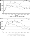
 ), A05830 (
 ), and A05898 (
 ) caused accelerated loss of mean fluorescence in KSHV-BJAB cultures as compared with the DMSO control (
 ), eventually establishing a new plateau level of fluorescence ≥50% less than the DMSO control. B, after incubation with extracts A05831 (
 ), A05853(
 ), and A05901 (
 ) fluorescence declined steadily and without leveling off, suggesting continuous reduction of the viral load.


Similar articles
-
Targeting human herpesvirus-8 for treatment of Kaposi's sarcoma and primary effusion lymphoma.Curr Opin Oncol. 2005 Sep;17(5):447-55. doi: 10.1097/01.cco.0000172823.01190.6c. Curr Opin Oncol. 2005. PMID: 16093794 Review.
-
Lytic induction of Kaposi's sarcoma-associated herpesvirus in primary effusion lymphoma cells with natural products identified by a cell-based fluorescence moderate-throughput screening.Arch Virol. 2008;153(8):1517-25. doi: 10.1007/s00705-008-0144-4. Epub 2008 Jul 8. Arch Virol. 2008. PMID: 18607675
-
Genipin Enhances Kaposi's Sarcoma-Associated Herpesvirus Genome Maintenance.PLoS One. 2016 Oct 13;11(10):e0163693. doi: 10.1371/journal.pone.0163693. eCollection 2016. PLoS One. 2016. PMID: 27736870 Free PMC article.
-
Kaposi's sarcoma-associated herpesvirus-encoded LANA contributes to viral latent replication by activating phosphorylation of survivin.J Virol. 2014 Apr;88(8):4204-17. doi: 10.1128/JVI.03855-13. Epub 2014 Jan 29. J Virol. 2014. PMID: 24478433 Free PMC article.
-
Viral latent proteins as targets for Kaposi's sarcoma and Kaposi's sarcoma-associated herpesvirus (KSHV/HHV-8) induced lymphoma.Curr Drug Targets Infect Disord. 2003 Jun;3(2):129-35. doi: 10.2174/1568005033481150. Curr Drug Targets Infect Disord. 2003. PMID: 12769790 Review.
Cited by
-
Targeting KSHV/HHV-8 latency with COX-2 selective inhibitor nimesulide: a potential chemotherapeutic modality for primary effusion lymphoma.PLoS One. 2011;6(9):e24379. doi: 10.1371/journal.pone.0024379. Epub 2011 Sep 30. PLoS One. 2011. PMID: 21980345 Free PMC article.
-
Update on HHV-8-Associated Malignancies.Curr Infect Dis Rep. 2010 Mar;12(2):147-54. doi: 10.1007/s11908-010-0092-5. Epub 2010 Mar 26. Curr Infect Dis Rep. 2010. PMID: 20461118 Free PMC article.
-
Glutamate secretion and metabotropic glutamate receptor 1 expression during Kaposi's sarcoma-associated herpesvirus infection promotes cell proliferation.PLoS Pathog. 2014 Oct 9;10(10):e1004389. doi: 10.1371/journal.ppat.1004389. eCollection 2014 Oct. PLoS Pathog. 2014. PMID: 25299066 Free PMC article.
-
Kaposi's sarcoma-associated herpesvirus latency in endothelial and B cells activates gamma interferon-inducible protein 16-mediated inflammasomes.J Virol. 2013 Apr;87(8):4417-31. doi: 10.1128/JVI.03282-12. Epub 2013 Feb 6. J Virol. 2013. PMID: 23388709 Free PMC article.
-
The open chromatin landscape of Kaposi's sarcoma-associated herpesvirus.J Virol. 2013 Nov;87(21):11831-42. doi: 10.1128/JVI.01685-13. Epub 2013 Aug 28. J Virol. 2013. PMID: 23986576 Free PMC article.
References
-
- Antman K, Chang Y. Kaposi's sarcoma. N Engl J Med. 2000;342:1027–38. - PubMed
-
- Cesarman E, Chang Y, Moore PS, Said JW, Knowles DM. Kaposi's sarcoma-associated herpesvirus-like DNA sequences in AIDS-related body-cavity-based lymphomas. N Engl J Med. 1995;332:1186–91. - PubMed
-
- Soulier J, Grollet L, Oksenhendler E, et al. Kaposi's sarcoma-associated herpesvirus-like DNA sequences in multicentric Castleman's disease. Blood. 1995;86:1276–80. - PubMed
-
- Harwood AR, Osoba D, Hofstader SL, et al. Kaposi's sarcoma in recipients of renal transplants. Am J Med. 1979;67:759–65. - PubMed
-
- Nador RG, Cesarman E, Chadburn A, et al. Primary effusion lymphoma: a distinct clinicopathologic entity associated with the Kaposi's sarcoma-associated herpes virus. Blood. 1996;88:645–56. - PubMed
Publication types
MeSH terms
Substances
Grants and funding
- HL083469/HL/NHLBI NIH HHS/United States
- CA096500/CA/NCI NIH HHS/United States
- U19 CA052956/CA/NCI NIH HHS/United States
- 5T32AI007419/AI/NIAID NIH HHS/United States
- R01 DE018304/DE/NIDCR NIH HHS/United States
- DE018281/DE/NIDCR NIH HHS/United States
- CA109232/CA/NCI NIH HHS/United States
- T32 CA071341/CA/NCI NIH HHS/United States
- 2P30AI050410/AI/NIAID NIH HHS/United States
- R01 HL083469/HL/NHLBI NIH HHS/United States
- R01 CA109232/CA/NCI NIH HHS/United States
- R01 CA163217/CA/NCI NIH HHS/United States
- 2T32CA071341/CA/NCI NIH HHS/United States
- DE018304/DE/NIDCR NIH HHS/United States
- T32 AI007419/AI/NIAID NIH HHS/United States
- R01 DE018281/DE/NIDCR NIH HHS/United States
- U19-CA52956/CA/NCI NIH HHS/United States
- P30 AI050410/AI/NIAID NIH HHS/United States
- R01 CA096500/CA/NCI NIH HHS/United States
LinkOut - more resources
Full Text Sources
Molecular Biology Databases

