. 2007 Aug 15;21(16):2055-68.
doi: 10.1101/gad.432007.

Angiomotin regulates endothelial cell migration during embryonic angiogenesis

Affiliations

Angiomotin regulates endothelial cell migration during embryonic angiogenesis

Karin Aase et al. Genes Dev. .

Abstract

The development of the embryonic vascular system into a highly ordered network requires precise control over the migration and branching of endothelial cells (ECs). We have previously identified angiomotin (Amot) as a receptor for the angiogenesis inhibitor angiostatin. Furthermore, DNA vaccination targeting Amot inhibits angiogenesis and tumor growth. However, little is known regarding the role of Amot in physiological angiogenesis. We therefore investigated the role of Amot in embryonic neovascularization during zebrafish and mouse embryogenesis. Here we report that knockdown of Amot in zebrafish reduced the number of filopodia of endothelial tip cells and severely impaired the migration of intersegmental vessels. We further show that 75% of Amot knockout mice die between embryonic day 11 (E11) and E11.5 and exhibit severe vascular insufficiency in the intersomitic region as well as dilated vessels in the brain. Furthermore, using ECs differentiated from embryonic stem (ES) cells, we demonstrate that Amot-deficient cells have intact response to vascular endothelial growth factor (VEGF) in regard to differentiation and proliferation. However, the chemotactic response to VEGF was abolished in Amot-deficient cells. We provide evidence that Amot is important for endothelial polarization during migration and that Amot controls Rac1 activity in endothelial and epithelial cells. Our data demonstrate a critical role for Amot during vascular patterning and endothelial polarization.


Figures

Figure 1.
Figure 1.
Amot is essential for ISV formation. Depletion of Amot expression in developing zebrafish embryos using amot antisense morpholinos leads to vascular defects in the head and trunk regions. (A–D) Lateral views; dorsal is up, anterior is to the left. The cranial arteries are normal in embryos injected with the mismatch control morpholino (A), whereas the pMBC and pHBC are dilated in the AmotKD embryos (B, arrowheads) at 36 hpf. The sprouting of the ISVs and the formation of the DLAV is complete at 36 hpf in the control (C), but the migration of the ECs in the ISVs are halted midway in the AmotKD (D, arrows), and no continuous SLAV is formed. (E) The percentage of defected embryos at 36 and 60 hpf using different morpholinos. Injection of amot antisense morpholinos leads to 65% defective embryos at 36 and 60 hpf, whereas the control mismatch embryos do not display any defects. Coinjection of human amot mRNA leads to rescue of the phenotypes. Coinjection of murine amotl-1 mRNA leads to a delayed rescue at 60 hpf. (F) The number of filopodia per cell was reduced fivefold in the AmotKD. AmotKD embryos display dilated pMBC and pHBC, and arrested ISV migration at 36 hpf. At 60 hpf, AmotKD embryos display only the ISV defects. Bar, 100 μm.
Figure 2.
Figure 2.
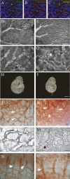
Vascular defects in the Amot-deficient yolk sacs and embryos at E10.5. Immunofluorescent staining at E10.5 using Amot-specific antibodies in combination with the endothelial marker isolectin-B4. Amot is expressed by the capillaries of the brain (A) where the staining overlaps with isolectin, a marker for EC (A–C). More expression data are found in Supplementary Figures 1 and 2. Wild-type yolk sacs form a branched vascular network with different sized vessels (D), whereas the Amot-deficient yolk sacs display a less-organized vessel network (E). At a higher magnification, the difference is shown more clearly, with narrow blood vessels in the wild-type (F) and dilated blood vessels that have anastomosed into lagoon-like structures (asterisk) in the Amot yolk sacs (G). Stage-matched E10.5 embryos of wild type (H) and Amot (I). (J) PECAM staining of the blood vessels in the brain show that in wild-type embryos, the blood vessels have expanded into a vascular plexus with large vessels that branch into smaller capillaries in the developing brain and somites (arrows). (K) The brains of the Amot embryos show similar structures as in the yolk sacs with dilated vessels (arrows) and lagoon-like structures (asterisk). There is an accumulation of erythrocytes in the Amot vessels (M), compared with the wild-type vessels (L). There is a lack of paraneural capillaries in the somitic region in the Amot embryos (O, arrowhead) that are present in the wild-type embryos (N, arrowheads). Bars: A–C, 14 μm; D,E,H,I, 1 mm; F,G, 21 μm; J–O, 10 μm.
Figure 3.
Figure 3.
VEGF-induced sprouting is impaired in Amot-deficient embryoid bodies. ES cells were allowed to differentiate and grow in three-dimensional collagen I gels. ECs were visualized with the PECAM antibody (red) and supporting perivascular cells with α-smooth muscle actin (αSMA) (green). (A) In the presence of VEGF, the wild-type cells migrate and form sprouting tubes that invade the collagen gel, whereas in the Amot cells only a few sprouts are formed. (B) Quantification of the tubes that invade the collagen I gel. (C) Invading tubes were coated with perivascular cells in both the wild-type and Amot bodies. (D) αSMA-positive cells were not dependent on either VEGF or Amot for their migration. Bars: A, 500 μm; B, 50 μm; C, 250 μm.
Figure 4.
Figure 4.
siRNA knockdown of Amot in the CNV model impairs angiogenic sprouting in vivo. Neovascularization was induced by laser-induced rupture of the innermost layer of the choroid. The lesions were treated with intraocular injections of siRNA at 0, 3, and 6 d. The sprouting ECs within the lesion were visualized using the PECAM antibody. Mice treated with the control siRNA (A) display extensive sprouting in the lesion, whereas the sprouting area of the lesion in mice treated with the Amot siRNA (B) is half the size (C). (***) P < 0.001.
Figure 5.
Figure 5.
Amot is not required for tight junction formation, but for the organization of actin and focal adhesions. (A) Wild-type PmT-ECs express both isoforms of Amot, whereas no expression is detected in the Amot PmT-ECs. (B) RT–PCR analysis shows that both cell lines express an equal amount of EC-specific markers. (C, top panel) Amot expression overlaps with the tight junction marker ZO-1 in cell–cell contacts in the wild-type PmT-EC. (Bottom panel) ZO-1 still localizes to cell–cell junctions in the absence of Amot. (D) Visualization of focal adhesions (arrowheads) and actin fibers using a paxillin antibody and phalloidin. (Bottom panel) The Amot PmT-EC displays an irregular pattern of actin fibers and shorter and smaller focal adhesions. The highlighted areas show focal adhesions at a higher magnification. Box diagram showing the area (E) and length (F) of focal adhesions. Bar, 14 μm. (***) P < 0.001.
Figure 6.
Figure 6.
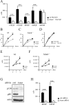
Amot is critical for growth factor-mediated migration. (A) Wild-type and Amot PmT-ECs were allowed to migrate toward VEGF, bFGF, or serum in the Boyden chamber assay. Amot PmT-ECs fail to respond to growth factors but show an equal migratory capacity as wild-type PmT-ECs in the presence of serum. In the in vitro wound healing assay, wild-type PmT-ECs respond to VEGF with an increase in migration rate (B), whereas the Amot PmT-ECs fail to respond (C). (D) There is no difference in migratory response in the presence of serum. (E,F) Both wild-type and Amot PmT-ECs display a similar response in proliferation to VEGF. (G) siRNA knockdown of Amot in BCE cells was confirmed using Western blot. (H) Amot siRNA-transfected BCE cells show a decrease in basal migration in the Boyden chamber assay and do not respond to bFGF.
Figure 7.
Figure 7.
Amot deficiency results in an increase in lamellipodia formation, an increase in Rac activity, and loss of polarization. (A, top panel) Photographs from time-lapse studies at indicated time points show that the wild-type PmT-ECs form one large lamellipodia (asterisk) in the direction of migration. (Bottom panel) In contrast, the Amot PmT-ECs form several cell protrusions and lamellipodia (arrows). (B) Subconfluent cells are stained with a Golgi apparatus-specific antibody and scored for degree of polarization. (C) A majority of the wild-type cells are polarized, whereas only about half of the Amot cells display the same pattern. Lamellipodia are highlighted with white dotted lines in A. Bar, 14 μm. (D) Rac1 activation is increased in the Amot PmT-ECs. (E) Knockdown of both isoforms of Amot in 293T cells also leads to an increase in the activation of Rac1. (F) Equal expression of Rich-1 in wild-type and Amot PmT-ECs. (G) In wild-type cells, Rich-1 localizes to lamellipodia (arrow, top panel), but its localization is altered in the Amot PmT-ECs (bottom panel).

Similar articles

Cited by

References

    1. Aepfelbacher M., Essler M., Huber E., Sugai M., Weber P.C., Essler M., Huber E., Sugai M., Weber P.C., Huber E., Sugai M., Weber P.C., Sugai M., Weber P.C., Weber P.C. Bacterial toxins block endothelial wound repair. Evidence that Rho GTPases control cytoskeletal rearrangements in migrating endothelial cells. Arterioscler. Thromb. Vasc. Biol. 1997;17:1623–1629. - PubMed
    1. Balconi G., Spagnuolo R., Dejana E., Spagnuolo R., Dejana E., Dejana E. Development of endothelial cell lines from embryonic stem cells: A tool for studying genetically manipulated endothelial cells in vitro. Arterioscler. Thromb. Vasc. Biol. 2000;20:1443–1451. - PubMed
    1. Berglin L., Sarman S., van der Ploeg I., Steen B., Ming Y., Itohara S., Seregard S., Kvanta A., Sarman S., van der Ploeg I., Steen B., Ming Y., Itohara S., Seregard S., Kvanta A., van der Ploeg I., Steen B., Ming Y., Itohara S., Seregard S., Kvanta A., Steen B., Ming Y., Itohara S., Seregard S., Kvanta A., Ming Y., Itohara S., Seregard S., Kvanta A., Itohara S., Seregard S., Kvanta A., Seregard S., Kvanta A., Kvanta A. Reduced choroidal neovascular membrane formation in matrix metalloproteinase-2-deficient mice. Invest. Ophthalmol. Vis. Sci. 2003;44:403–408. - PubMed
    1. Bostrom H., Willetts K., Pekny M., Leveen P., Lindahl P., Hedstrand H., Pekna M., Hellstrom M., Gebre-Medhin S., Schalling M., Willetts K., Pekny M., Leveen P., Lindahl P., Hedstrand H., Pekna M., Hellstrom M., Gebre-Medhin S., Schalling M., Pekny M., Leveen P., Lindahl P., Hedstrand H., Pekna M., Hellstrom M., Gebre-Medhin S., Schalling M., Leveen P., Lindahl P., Hedstrand H., Pekna M., Hellstrom M., Gebre-Medhin S., Schalling M., Lindahl P., Hedstrand H., Pekna M., Hellstrom M., Gebre-Medhin S., Schalling M., Hedstrand H., Pekna M., Hellstrom M., Gebre-Medhin S., Schalling M., Pekna M., Hellstrom M., Gebre-Medhin S., Schalling M., Hellstrom M., Gebre-Medhin S., Schalling M., Gebre-Medhin S., Schalling M., Schalling M., et al. PDGF-A signaling is a critical event in lung alveolar myofibroblast development and alveogenesis. Cell. 1996;85:863–873. - PubMed
    1. Bratt A., Wilson W.J., Troyanovsky B., Aase K., Kessler R., Van Meir E.G., Holmgren L., Wilson W.J., Troyanovsky B., Aase K., Kessler R., Van Meir E.G., Holmgren L., Troyanovsky B., Aase K., Kessler R., Van Meir E.G., Holmgren L., Aase K., Kessler R., Van Meir E.G., Holmgren L., Kessler R., Van Meir E.G., Holmgren L., Van Meir E.G., Holmgren L., Holmgren L. Angiomotin belongs to a novel protein family with conserved coiled-coil and PDZ binding domains. Gene. 2002;298:69–77. - PubMed

Publication types

MeSH terms