

Brain MRI findings in infants with primary congenital glaucoma
- PMID: 17700055
- PMCID: PMC6074287
- DOI: 10.5144/0256-4947.2007.264
Brain MRI findings in infants with primary congenital glaucoma
Abstract
Background: Congenital glaucoma appears in the first months of life, eventually at birth. Isolated congenital glaucoma is characterized by minor malformations of the irido-corneal angle of the anterior chamber of the eye. Clinical manifestations include tearing, photophobia and enlargement of the globe appearing in the first months of life. Imaging technology such as optical coherence tomography and measurement of central corneal thickness may play an important role in the assessment of children with suspected or known glaucoma. However, no MRI findings of the CNS in patients with primary congenital glaucoma (PCG) were reported in the literature. The purpose of this study was to investigate MRI findings of the brain in infants with PCG.
Methods: We reviewed the radiological, histopathological and clinical characteristics of infants with primary congenital glaucoma. The records of 17 patients with PCG were reviewed and the MRIs of the brain and associated manifestations were analyzed.
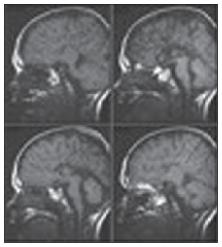
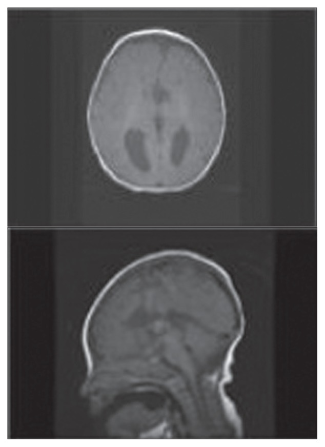
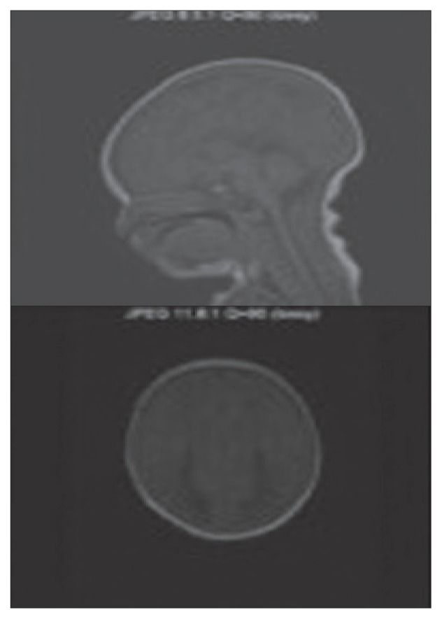
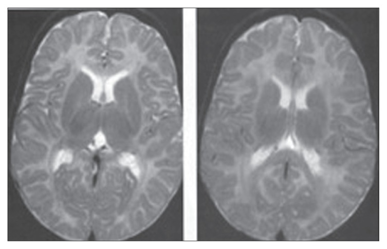
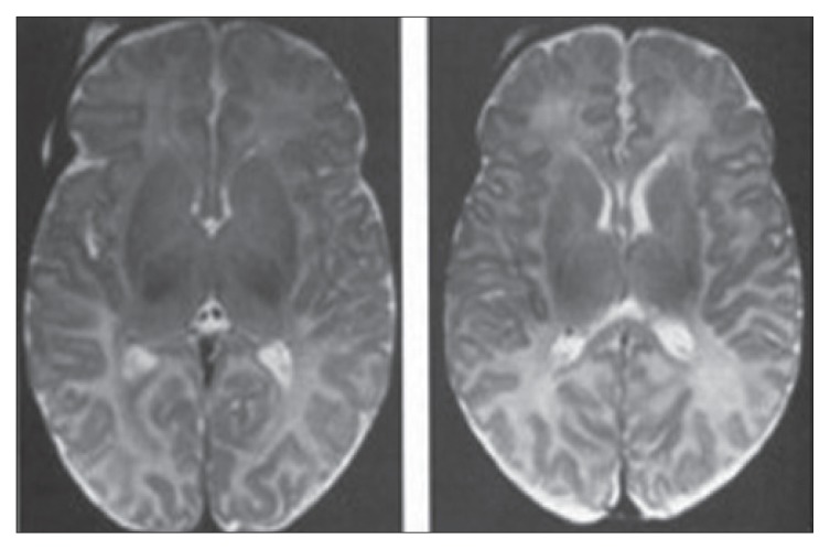
Results: Three patients with PCG had abnormal MRI findings suggesting agenesis of the corpus callosum. Two infants had delayed myelinization of the brain.
Discussion: Significant abnormal optic nerve excavation and increased corneal diameters in 2 patients with delayed myelinization may suggest that intraocular pressure can be more striking and more severe, revealing a close relationship with PCG and abnormal myelinization in the white matter. Studies with more patients are needed to confirm these results.
Figures
Similar articles
-
Rare Case Of Primary Congenital Glaucoma With Hypoplasia Corpus Callosum.J Ayub Med Coll Abbottabad. 2018 Apr-Jun;30(2):286-288. J Ayub Med Coll Abbottabad. 2018. PMID: 29938437
-
Spectral domain optical coherence tomography in children operated for primary congenital glaucoma.Br J Ophthalmol. 2014 Feb;98(2):162-5. doi: 10.1136/bjophthalmol-2012-302486. Epub 2013 Jun 5. Br J Ophthalmol. 2014. PMID: 23740961
-
[Primary congenital glaucoma].Harefuah. 2004 Dec;143(12):876-80, 910. Harefuah. 2004. PMID: 15666707 Review. Hebrew.
-
Clinical features and neuroimaging (CT and MRI) findings in presumed Zika virus related congenital infection and microcephaly: retrospective case series study.BMJ. 2016 Apr 13;353:i1901. doi: 10.1136/bmj.i1901. BMJ. 2016. PMID: 27075009 Free PMC article.
-
Primary congenital glaucoma.Prog Brain Res. 2015;221:177-89. doi: 10.1016/bs.pbr.2015.06.005. Epub 2015 Sep 9. Prog Brain Res. 2015. PMID: 26518078 Review.
Cited by
-
Advanced Diffusion MRI of the Visual System in Glaucoma: From Experimental Animal Models to Humans.Biology (Basel). 2022 Mar 16;11(3):454. doi: 10.3390/biology11030454. Biology (Basel). 2022. PMID: 35336827 Free PMC article. Review.
-
Computed Tomographic Thickness of Retrobulbar Optic Nerve Is Decreased in Glaucoma Patients and Is Negatively Correlated With Disease Severity.Cureus. 2022 Nov 3;14(11):e31066. doi: 10.7759/cureus.31066. eCollection 2022 Nov. Cureus. 2022. PMID: 36475192 Free PMC article.
References
-
- Ho CL, Walton DS. Primary congenital glaucoma: 2004 update. J Pediatr Ophthalmol Strabısmus. 2004;41:271–88. - PubMed
-
- Mandal AK, Gothwal VK, Bagga H, Nutheti R. Outcome of Surgery on Infants Younger than 1 Month with Congenital Glaucoma. Ophthalmology. 2003;110:1909–15. - PubMed
-
- Akpek EK, Jun AS, Goodman DF, Green WR, Gottsch JD. Clinical and Ultrastructural Features of a Novel Hereditary Anterior Segment Dysgenesis. Ophthalmology. 2002;109:513–519. - PubMed
-
- The AGIS investigators. The advanced glaucoma intervention study (AGIS). The relationship between control of intraocular pressure and visual field deterioration. Am J Ophthalmol. 2000;130:429–440. - PubMed
-
- Heijl MC, Leske B, Bengtsson L, Hyman B, Bengtsson M Early Manifest Glaucoma Trial Group. Reduction of intraocular pressure and glaucoma progression. Results from the early manifest glaucoma trial, Arch Ophthalmol. 2002;120:1268–1279. - PubMed
MeSH terms
LinkOut - more resources
Full Text Sources
Medical