

Molecular analysis in combination with iodine staining may contribute to the risk prediction of esophageal squamous cell carcinoma
- PMID: 17701052
- PMCID: PMC12161663
- DOI: 10.1007/s00432-007-0269-y
Molecular analysis in combination with iodine staining may contribute to the risk prediction of esophageal squamous cell carcinoma
Abstract
Purpose: Mucosal iodine staining has improved the detection of precancerous lesions of the esophagus. However, this method is unable to exactly evaluate the risk status of the lesions. In the present study, we conducted a molecular analysis combining the iodine staining in esophageal squamous cell carcinomas (ESCC) and different premalignant lesions of the esophagus in order to improve the early diagnosis of ESCC.
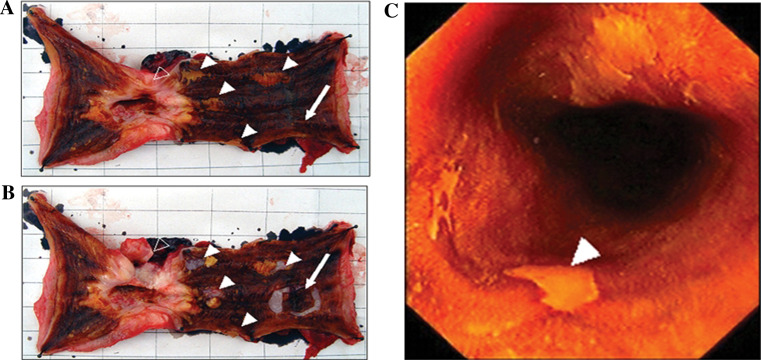
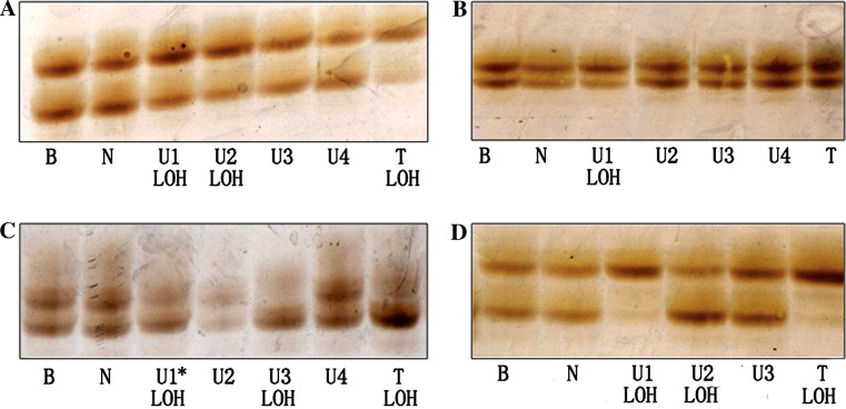
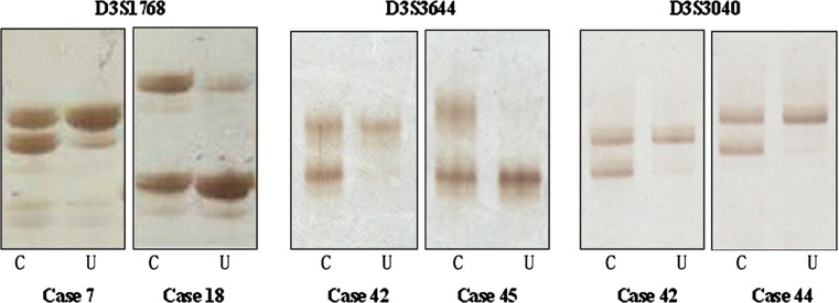
Methods: Tumorous and precancerous lesions were procured as iodine-unstained areas in the resected specimens of ESCC patients by means of Lugol's iodine staining. Loss of heterozygosity (LOH) was detected with 35 microsatellite markers frequently reported to be deleted in ESCC. The markers with high frequency of LOH in tumorous and precancerous lesions of the same patient were subjected to further detection in iodine-unstained biopsy samples from the population screening in ESCC high-incidence region.
Results: Common alterations were observed at D3S3644, D3S1768, D3S3040, D3S4542, RPL14, D9S169, D13S171 and D13S263 in both cancer tissues and precancerous lesions around tumors. Interestingly, D3S3644, D3S1768, D3S3040, D3S4542, RPL14 and D13S263 were also found with high frequency of LOH in iodine-staining abnormal lesions from the population screening. Most importantly, LOH frequency increased with histological severity.
Conclusion: Our data suggest that detection of these six markers in combination with iodine staining might contribute to the prediction for the risk of ESCC development and for the diagnosis of patients in preclinical and preneoplastic phase of the disease.
Figures
References
-
- Braakhuis BJ, Tabor MP, Kummer JA, Leemans CR, Brakenhoff RH (2003) A genetic explanation of Slaughter’s concept of field cancerization: evidence and clinical implications. Cancer Res 63:1727–1730 - PubMed
-
- Dawsey SM, Lewin KJ, Liu FS, Wang GQ, Shen Q (1994a) Esophageal morphology from Linxian, China. Squamous histologic findings in 754 patients. Cancer 73:2027–2037 - PubMed
-
- Dawsey SM, Lewin KJ, Wang GQ, Liu FS, Nieberg RK, Yu Y, Li JY, Blot WJ, Li B, Taylor PR (1994b) Squamous esophageal histology and subsequent risk of squamous cell carcinoma of the esophagus. A prospective follow-up study from Linxian, China. Cancer 74:1686–1692 - PubMed
-
- Dawsey SM, Fleischer DE, Wang GQ, Zhou B, Kidwell JA, Lu N, Lewin KJ, Roth MJ, Tio TL, Taylor PR (1998) Mucosal iodine staining improves endoscopic visualization of squamous dysplasia and squamous cell carcinoma of the esophagus in Linxian, China. Cancer 83:220–231 - PubMed
-
- Dong Z, Tang P, Li L, Wang G (2002) The strategy for esophageal cancer control in high-risk areas of China. Jpn J Clin Oncol 32(Suppl):S10–12 - PubMed
Publication types
MeSH terms
Substances
LinkOut - more resources
Full Text Sources
Medical
Miscellaneous
