. 2007 Oct 1;37(4):1161-77.
doi: 10.1016/j.neuroimage.2007.05.020. Epub 2007 Jul 4.

Robust detection of ocular dominance columns in humans using Hahn Spin Echo BOLD functional MRI at 7 Tesla

Affiliations

Robust detection of ocular dominance columns in humans using Hahn Spin Echo BOLD functional MRI at 7 Tesla

Essa Yacoub et al. Neuroimage. .

Abstract

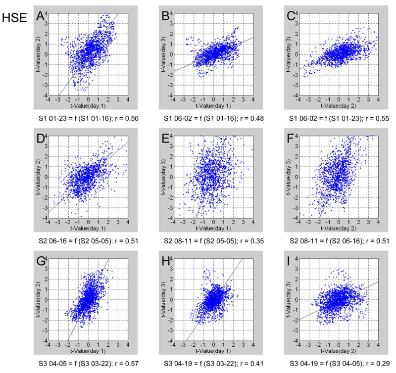
Cells in the mammalian brain tend to be grouped together according to their afferent and efferent connectivity, as well as their physiological properties. The columnar structures of neocortex are prominent examples of such modular organization, and have been studied extensively in anatomical and physiological experiments in rats, cats and monkeys. The importance of noninvasive study of such structures, in particular in human subjects, cannot be overemphasized. Not surprisingly, therefore, many attempts were made to map cortical columns using functional magnetic resonance imaging (fMRI). Yet, the robustness, repeatability, and generality of the hitherto used fMRI methodologies have been a subject of intensive debate. Using differential mapping in a high magnetic field magnet (7 T), we demonstrate here the ability of Hahn Spin-Echo (HSE) BOLD to map the ocular dominance columns (ODCs) of the human visual cortex reproducibly over several days with a high degree of accuracy, relative to expected spatial patterns from post-mortem data. On the other hand, the conventional Gradient-Echo (GE) blood oxygen level dependent (BOLD) signal in some cases failed to resolve ODCs uniformly across the selected gray matter region, due to the presence of non-specific signals. HSE signals uniformly resolved the ODC patterns, providing a more generalized mapping methodology (i.e. one that does not require adjusting experimental approaches based on prior knowledge or assumptions about functional organization and vascular structure in order to avoid confounding large vessel effects) to map unknown columnar systems in the human brain, potentially paving the way both for the study of the functional architecture of human sensory cortices, and of brain modules underlying specific cognitive processes.


Figures

Figure 1
Figure 1
An example of slice selection from subject 3. The slice was optimized for covering the flat portion of cortex within the lower bank of the calcarine fissure in the right hemisphere (RH). The ODCs from the right hemisphere are expected to run within the slice orthogonally to the midline.
Figure 2
Figure 2
An example of matching slices obtained on different days from subject 3. The 3 images depict the imaging slice selected from the same subject on 3 different days. The images from day 2 and 3 were registered to the image from day 1 (the image to the left). The red arrows point to the same landmarks as they appear on the 3 different images. The green box indicates the flat gray matter region within the calcarine fissure that was selected as the ROI.
Figure 3
Figure 3
Differential functional OD maps depicting increased activity for left eye stimulation (blue) and right eye stimulation (red) for Subject 1. The upper and lower rows depict the maps obtained using GE and HSE fMRI, respectively. (A–C) OD maps from 3 different GE sessions. These maps were obtained after registering the functional data from day 2 (B) and day 3 (C) to the data from day 1 (A). (D–E) The overlap between maps obtained in the 2 most reproducible GE sessions (D) and all 3 (E) of these GE sessions. For computing the maps presented in D–E, the differential maps obtained in different sessions were spatially filtered (see methods). A logical AND operator was applied to the maps obtained on different days. In D, the maps obtained in days 1 and 3 (presented in A and C) are compared. Red or blue colored voxels passed the threshold on the 2 (D) or all 3 (E) days, and showed consistent eye preference over days. The bottom row depicts in an identical format the maps obtained from 3 different HSE sessions with registered data (F–H), and the overlap between maps obtained in 2 (I) and all 3 (J) HSE sessions. In I, the maps obtained in days 1 and 2 (presented in F and G) are compared. The registrations of GE data (A–E) and HSE data (F–J) were performed separately. The superimposed arrows were positioned according to the HSE overlap map shown in (I), and copied to locations that are identical relative to the registered HSE data (F–J). Therefore they can facilitate the evaluation of reproducibility. The same set of arrows was superimposed onto the registered GE maps corresponding to the approximate locations that were determined according to anatomical landmarks and the GE overlap map shown in (D). The same statistical threshold was used for all studies (p < .15). The background image in A–C and F–H are the BOLD GE and HSE images, respectively. The background gray level images in D–E and I–J show the differential OD response averaged over the compared 2 or 3 sessions. Pos, posterior; RH, right hemisphere, IHF, Inter-hemispheric fissure.
Figure 4
Figure 4
Differential OD maps obtained from Subject 2. The upper and lower rows depict the maps obtained using GE and HSE fMRI, respectively. The analysis procedure and the format of presentation are identical to those used in Fig. 3. In D, the maps obtained in days 1 and 3 (presented in A and C) are compared. In I, the maps obtained in days 1 and 2 (presented in F and G) are compared.
Figure 5
Figure 5
Differential OD maps obtained from Subject 3. The upper and lower rows depict the maps obtained using GE and HSE fMRI, respectively. The analysis procedure and format of presentation are identical to those used in Fig. 3. In D, the maps obtained in days 1 and 2 (presented in A and B) are compared. In I, the maps obtained in days 1 and 2 (presented in F and G) are compared.
Figure 6
Figure 6
Reproducibility across days of ODC maps obtained using GE BOLD signals. Plots show the scatter of t-values of individual voxels from one scan day versus the t-values of the corresponding voxels from a second scan day. The data from each pair of compared days were spatially aligned. The first, second, and third row presents comparisons of imaging sessions of subject 1, 2, and 3, respectively. All voxels which passed the differential statistical threshold, irrespective of any column-like patterns were used in the analysis. Voxels that fall in the upper right quadrant demonstrated preference for right eye stimulation on both days. Voxels in the lower left quadrant demonstrated preference for left eye stimulation on both days. The preference for eye stimulation of voxels in the other quadrants was not reproducible over the 2 sessions. The straight line depicts regression using orthogonal fitting.
Figure 7
Figure 7
Reproducibility across days of ODC maps obtained using HSE BOLD signals. The format of presentation is identical to that in Figure 6.
Figure 8
Figure 8
Estimation of false positive ODC detection: functional maps depicting differential analysis of HSE data obtained during binocular stimulation. (A) and (B) depict the results of 2 different sessions from subject 2. The data were registered to the HSE data presented in Fig. 4. The arrows were copied from Fig. 4 and registered to the same anatomical landmarks. The activated voxels here are a measure of false positive detection resulting from the data acquisition and analysis procedures we used. (C) The overlap between maps presented in (A) and (B). The differential maps obtained in different sessions were spatially filtered (see methods). A logical AND operator was applied to the maps obtained on different days, in a manner similar to that used for the reproducibility map presented in Fig. 4I. (D) Reproducibility across days of the differential t-values computed using differential analysis of HSE data obtained during binocular stimulation. The data used for the map shown in B is presented as a function of the data used for the map in A. The data from the two compared sessions were spatially registered prior to the analysis. The format of presentation is identical to that in Figs. 6–7. In contrast to the reproducibility shown in Fig. 7, the scatter plot shows a random relationship here. (E) The data used for the map shown in A (with binocular stimulation) is presented as a function of the data used for the map in Fig. 4F (with monocular stimulation). (F) The data used for the map shown in B (with binocular stimulation) is presented as a function of the data used for the map in Fig. 4F (with monocular stimulation).
Figure 9
Figure 9
Spatial frequency distribution of the differential OD maps. Each of the presented distributions is the average over the 3 spatial frequency distributions obtained in 3 sessions from one subject. HSE BOLD and GE BOLD maps are shown at the top and bottom row, respectively. The data from Subject 1 (left) showed clear peaks approximately at the expected orientation (above and below the center, representing the anterior-posterior axis) and spatial frequency (0.5 cycles / mm) of OD maps, for both GE and HSE data. The data from Subject 2 (center) showed clear peaks at the expected locations for the HSE BOLD data. In contrast, no increased power was observed in those locations for the GE data. The data from subject 3 showed a clearer increase in power at the expected locations for the HSE data than for the GE data. The arrows indicate the locations of the observed increase in power in frequencies approximately expected for ODCs (~ 0.5 cycles / mm, oscillating along the anterior-posterior axis).
Figure 10
Figure 10
BOLD response relative to baseline from subject 1 and subject 3, respectively, for GE and HSE data from ODCs showing preference to the stimulated (labeled ‘STIMULATED’) and the non-stimulated (labeled ‘NON-STIMULATED’) eye. The response was measured in similar regions where both GE and HSE BOLD data showed ODC patterns. Error bars represent the SD between the different scan days. Differential response: the response from ODCs with preference to the non-stimulated eye was subtracted from the response measured at ODCs with preference to the stimulated eye. Selectivity index: differential contrast relative to the global response from baseline ((% change in ODCs with preference to the stimulated eye - % change in ODCs with preference to the non-stimulated eye)/ % change in ODCs with preference to the stimulated eye). The mean selectivity index was 0.52 ± 0.005 for HSE data and 0.24 ± 0.13 for GE data.

References

    1. Adriany G, Pfeuffer J, Yacoub E, Van de Moortele P-F, Shmuel A, Andersen P, Hu X, Vaughan JT, Ugurbil K. A half-volume transmit/ receive coil combination for 7 Tesla applications. ISMRM; Glasgow, U.K: 2001. p. 1097.
    1. Albright TD, Desimone R, Gross CG. Columnar organization of directionally selective cells in visual area MT of the macaque. J Neurophysiol. 1984;51:16–31. - PubMed
    1. Blasdel GG. Differential imaging of ocular dominance and orientation selectivity in monkey striate cortex. J Neurosci. 1992;12:3115–3138. - PMC - PubMed
    1. Blasdel GG, Salama G. Voltage-sensitive dyes reveal a modular organization in monkey striate cortex. Nature. 1986;321:579–585. - PubMed
    1. Bonhoeffer T, Grinvald A. Iso-orientation domains in cat visual cortex are arranged in pinwheel-like patterns. Nature. 1991;353:429–431. - PubMed

Publication types