

Review
doi: 10.1016/j.semcdb.2007.06.008.
Epub 2007 Jul 10.
Membrane type 1-matrix metalloproteinase: substrate diversity in pericellular proteolysis
Affiliations
- PMID: 17702616
- PMCID: PMC2685078
- DOI: 10.1016/j.semcdb.2007.06.008
Item in Clipboard
Review
Membrane type 1-matrix metalloproteinase: substrate diversity in pericellular proteolysis
Semin Cell Dev Biol.
2008 Feb.
Abstract
Enzymes in the matrix metalloproteinase (MMP) family have been linked to key events in developmental biology for almost 50 years. Biochemical, cellular and in vivo analyses have established that pericellular proteolysis contributes to numerous aspects of ontogeny including ovulation, fertilization, implantation, cellular migration, tissue remodeling and repair. Surface anchoring of proteinase activity provides spatial restrictions on substrate targeting. This review will utilize membrane type 1 MMP (MT1-MMP) as an example to highlight substrate diversity in pericellular proteolysis catalyzed by a membrane anchored MMP.
Figures
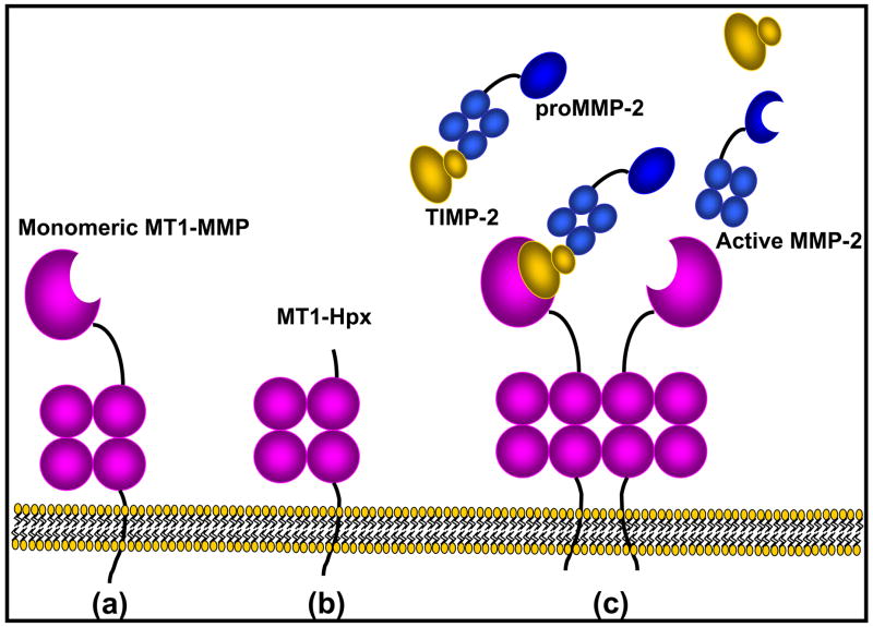
(a) Mature membrane-anchored MT1-MMP is comprised of a catalytic domain (Y-G285) containing the Zn++-binding consensus sequence, a hinge region (E286-I318), hemopexin-like domain (C319-C508), a membrane-adjacent stalk region (P509-S538), a transmembrane domain (A539-F562) and a cytoplasmic tail (R563-V582). (b) MT1-MMP undergoes autolytic processing at G284-G285 to generate a membrane-anchored species lacking the catalytic domain but retaining a surface-localized hemopexin domain that regulates activity of the mature enzyme. (c) MT1-MMP-catalyzed activation of proMMP-2 involves two molecules of MT1-MMP, likely functioning as a dimeric unit. One member of the complex forms a trimeric activation complex comprised of MT1-MMP, TIMP-2 and proMMP-2. The proMMP-2 in the trimeric complex is properly positioned for efficient activation by TIMP-2-free MT1-MMP, generating active MMP-2.

(A, B) OvCa433 cells, with low endogenous MT1-MMP expression were transfected with either (A) a catalytically inactive (E240A mutant) or (B) wild type MT1-MMP and subcultured onto type I collagen gels containing quenched fluorescent type I collagen (DQ-collagen, Invitrogen). This collagen substrate is heavily conjugated with multiple fluorescein labels, leading to quenched fluorescence in the intact substrate. Upon hydrolysis to single dye labeled products, quenching is relieved yielding green fluorescent products indicative of collagenase activity. Note the pericellular collagenolysis in cells expressing wild type active MT1-MMP (B). (C) Invasion of type I collagen gels. OvCa433 cells transfected with wild type or catalytically inactive mutant (E240A) MT1-MMP or vector controls were seeded onto Transwell filters coated with a type I collagen gel and allowed to invade for 24 hr. Non-invading cells were removed from the upper chamber with a cotton swab prior to staining and enumeration of cells adherent to the underside of the filter using an ocular micrometer. Data from triplicate experiments are presented with S.D. value shown (*p<.005). (Figures courtesy of Yueying Liu.)
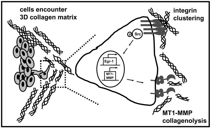
Migrating cells encountering a three-dimensional collagen matrix engage collagen via integrin-mediated attachments. Clustering of integrins resulting from interaction with 3-dimensional collagen induces phosphorylation of Src kinases, resulting in induction of the transcription factor Egr-1 and transcriptional activation of the MT1-MMP promoter. The active proteinase is trafficked to the cell surface where it participates in pericellular collagenolysis.

MT1-MMP catalyzes limited proteolytic processing and/or ectodomain shedding of a variety of membrane-anchored molecules including cadherins, integrins, proteoglycans (PG) and PG receptors as well as other surface substrates including LRP, RANKL, semaphoring 4D etc. These cleavages have been shown to regulate diverse cellular processes as summarized above.
Similar articles
-
MT1-MMP-dependent cell migration: proteolytic and non-proteolytic mechanisms.Biochem Soc Trans. 2019 Jun 28;47(3):811-826. doi: 10.1042/BST20180363. Epub 2019 May 7. Biochem Soc Trans. 2019. PMID: 31064864 Free PMC article. Review.
-
Matrix metalloproteinase-14 both sheds cell surface neuronal glial antigen 2 (NG2) proteoglycan on macrophages and governs the response to peripheral nerve injury.J Biol Chem. 2015 Feb 6;290(6):3693-707. doi: 10.1074/jbc.M114.603431. Epub 2014 Dec 8. J Biol Chem. 2015. PMID: 25488667 Free PMC article.
-
Identification and characterization of Lutheran blood group glycoprotein as a new substrate of membrane-type 1 matrix metalloproteinase 1 (MT1-MMP): a systemic whole cell analysis of MT1-MMP-associating proteins in A431 cells.J Biol Chem. 2009 Oct 2;284(40):27360-9. doi: 10.1074/jbc.M109.029124. Epub 2009 Aug 10. J Biol Chem. 2009. PMID: 19667067 Free PMC article.
-
Tetraspanin proteins regulate membrane type-1 matrix metalloproteinase-dependent pericellular proteolysis.Mol Biol Cell. 2009 Apr;20(7):2030-40. doi: 10.1091/mbc.e08-11-1149. Epub 2009 Feb 11. Mol Biol Cell. 2009. PMID: 19211836 Free PMC article.
-
MT1-MMP: a key regulator of cell migration in tissue.IUBMB Life. 2006 Oct;58(10):589-96. doi: 10.1080/15216540600962818. IUBMB Life. 2006. PMID: 17050376 Review.
Cited by
-
Non-destructive and selective imaging of the functionally active, pro-invasive membrane type-1 matrix metalloproteinase (MT1-MMP) enzyme in cancer cells.J Biol Chem. 2013 Jul 12;288(28):20568-80. doi: 10.1074/jbc.M113.471508. Epub 2013 Jun 3. J Biol Chem. 2013. PMID: 23733191 Free PMC article.
-
Proteolytic cleavage of membrane proteins by membrane type-1 MMP regulates cancer malignant progression.Cancer Sci. 2023 Feb;114(2):348-356. doi: 10.1111/cas.15638. Epub 2022 Nov 24. Cancer Sci. 2023. PMID: 36336966 Free PMC article. Review.
-
Structural basis for the sheddase function of human meprin β metalloproteinase at the plasma membrane.Proc Natl Acad Sci U S A. 2012 Oct 2;109(40):16131-6. doi: 10.1073/pnas.1211076109. Epub 2012 Sep 17. Proc Natl Acad Sci U S A. 2012. PMID: 22988105 Free PMC article.
-
A membrane-type-1 matrix metalloproteinase (MT1-MMP)-discoidin domain receptor 1 axis regulates collagen-induced apoptosis in breast cancer cells.PLoS One. 2015 Mar 16;10(3):e0116006. doi: 10.1371/journal.pone.0116006. eCollection 2015. PLoS One. 2015. PMID: 25774665 Free PMC article.
-
Physiology and pathophysiology of matrix metalloproteases.Amino Acids. 2011 Jul;41(2):271-90. doi: 10.1007/s00726-010-0689-x. Epub 2010 Jul 18. Amino Acids. 2011. PMID: 20640864 Free PMC article. Review.
References
-
- Ellerbroek SM, Stack MS. Membrane associated matrix metalloproteinases in metastasis. Bioessays. 1999;21(11):940–949. - PubMed
-
- Ellerbroek SM, Stack MS. Regulatory Mechanisms for Proteinase Activity. In: Simons HJSaC., editor. Proteinase and Peptidase Inhibition. Taylor and Francis; 2002. pp. 21–34.
-
- Werb Z. ECM and cell surface proteolysis: regulating cellular ecology. Cell. 1997;91(4):439–442. - PubMed
-
- Sato H, Takino T, Kinoshita T, et al. Cell surface binding and activation of gelatinase A induced by expression of membrane-type-1-matrix metalloproteinase (MT1-MMP) FEBS Letters. 1996;385(3):238–240. - PubMed
Publication types
MeSH terms
Substances
Grants and funding
LinkOut - more resources
Full Text Sources
